Polymer-Based 3D Printed Scaffolds for Tissue Engineering
S. Aslan, Y. Kaya, Y. Öztürk
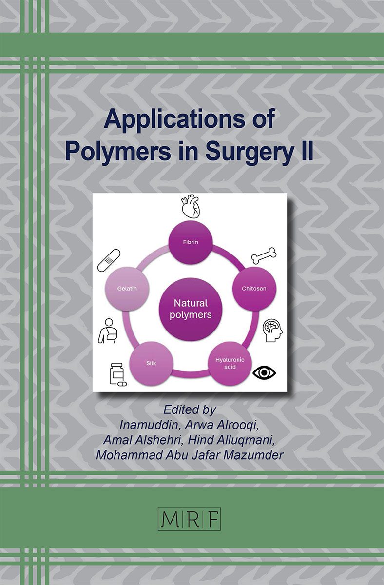
The way of 3D printing technology has diverged into many fields, with tissue engineering standing out as one of the most promising areas of application. Polymer-based 3D printed scaffolds are essential to tissue engineering because they offer structural support and facilitate cell growth and tissue regeneration. This review explores the advances in polymer-based 3D printed scaffolds, including materials used, fabrication techniques, and their applications in tissue engineering. Emphasis is placed on the properties of different polymers, the integration of bioactive factors, and the challenges and potential paths forward in this quickly developing subject.
Keywords
Polymers, 3D Scaffolds, Tissue Engineering, Technology
Published online 2/15/2025, 15 pages
Citation: S. Aslan, Y. Kaya, Y. Öztürk, Polymer-Based 3D Printed Scaffolds for Tissue Engineering, Materials Research Foundations, Vol. 172, pp 231-245, 2025
DOI: https://doi.org/10.21741/9781644903353-9
Part of the book on Applications of Polymers in Surgery II
References
[1] G. R. Strobl, The physics of polymers, vol. 2. Springer, 1997. https://doi.org/10.1007/978-3-662-03488-0
[2] S. C. Rasmussen, “Revisiting the early history of synthetic polymers: critiques and new insights,” Ambix, vol. 65, no. 4, pp. 356-372, 2018. https://doi.org/10.1080/00026980.2018.1512775
[3] G. Wypych, Handbook of polymers. Elsevier, 2022.
[4] O. Olatunji, Natural polymers: industry techniques and applications. Springer, 2015.
[5] K. Kaushik, R. B. Sharma, and S. Agarwal, “Natural polymers and their applications,” Int. J. Pharm. Sci. Rev. Res., vol. 37, no. 2, pp. 30-36, 2016.
[6] G. Satchanska, S. Davidova, and P. D. Petrov, “Natural and Synthetic Polymers for Biomedical and Environmental Applications,” Polymers (Basel)., vol. 16, no. 8, p. 1159, 2024. https://doi.org/10.3390/polym16081159
[7] M. F. Maitz, “Applications of synthetic polymers in clinical medicine,” Biosurface and Biotribology, vol. 1, no. 3, pp. 161-176, 2015. https://doi.org/10.1016/j.bsbt.2015.08.002
[8] M. C. Hacker, J. Krieghoff, and A. G. Mikos, “Synthetic polymers,” in Principles of regenerative medicine, Elsevier, 2019, pp. 559-590. https://doi.org/10.1016/B978-0-12-809880-6.00033-3
[9] S. Bhatia and S. Bhatia, “Natural polymers vs synthetic polymer,” Nat. Polym. drug Deliv. Syst. nanoparticles, plants, algae, pp. 95-118, 2016. https://doi.org/10.1007/978-3-319-41129-3_3
[10] L. E. Nielsen, “Cross-linking-effect on physical properties of polymers,” J. Macromol. Sci. Part C, vol. 3, no. 1, pp. 69-103, 1969. https://doi.org/10.1080/15583726908545897
[11] V. Ambrogi, C. Carfagna, P. Cerruti, and V. Marturano, “Additives in polymers,” in Modification of polymer properties, Elsevier, 2017, pp. 87-108. https://doi.org/10.1016/B978-0-323-44353-1.00004-X
[12] R. Geyer, “Production, use, and fate of synthetic polymers,” in Plastic waste and recycling, Elsevier, 2020, pp. 13-32. https://doi.org/10.1016/B978-0-12-817880-5.00002-5
[13] S. Ramakrishnan, “Condensation polymerization,” Resonance, vol. 22, pp. 355-368, 2017. https://doi.org/10.1007/s12045-017-0475-0
[14] L.-S. Hornberger, P. Weingarten, P. L. Lange, T. Schleid, and F. Adams, “Connecting 16- with 4-Membered Rings: Ring-Opening Polymerization of Bio-Based ω-Pentadecalactone with Amino-Alkoxy-Bis(phenolate) Yttrium Initiators and Copolymerization with β-Butyrolactone,” Eur. Polym. J., vol. 199, p. 112449, 2023. https://doi.org/10.1016/j.eurpolymj.2023.112449
[15] A. N. Generalova et al., “Polymers in 3D printing of external maxillofacial prostheses and in their retention systems,” Int. J. Pharm., vol. 657, p. 124181, 2024. https://doi.org/10.1016/j.ijpharm.2024.124181
[16] G. W. Ehrenstein and S. Pongratz, “Applications,” G. W. Ehrenstein and S. B. T.-R. and S. of P. Pongratz, Eds. Hanser, 2013, pp. 423-884. https://doi.org/10.1007/978-3-446-43709-8_5
[17] V. Mubayi, C. B. Ahern, M. Calusinska, and M. A. O’Malley, “Toward a Circular Bioeconomy: Designing Microbes and Polymers for Biodegradation,” ACS Synth. Biol., vol. 13, no. 7, pp. 1978-1993, 2024. https://doi.org/10.1021/acssynbio.4c00077
[18] J. E. Mark, Physical properties of polymers handbook. Newyork: Springer, 2007. https://doi.org/10.1007/978-0-387-69002-5
[19] T. Li, J. Chang, Y. Zhu, and C. Wu, “3D printing of bioinspired biomaterials for tissue regeneration,” Adv. Healthc. Mater., vol. 9, no. 23, p. 2000208, 2020. https://doi.org/10.1002/adhm.202000208
[20] M. S. Kim, J. H. Kim, B. H. Min, H. J. Chun, D. K. Han, and H. B. Lee, “Polymeric scaffolds for regenerative medicine,” Polym. Rev., vol. 51, no. 1, pp. 23-52, 2011. https://doi.org/10.1080/15583724.2010.537800
[21] R. Boni, A. Ali, A. Shavandi, and A. N. Clarkson, “Current and novel polymeric biomaterials for neural tissue engineering,” J. Biomed. Sci., vol. 25, pp. 1-21, 2018. https://doi.org/10.1186/s12929-018-0491-8
[22] J. Liu and C. Yan, “3D printing of scaffolds for tissue engineering,” by Cvetković D., Intech Open, UK, vol. 7, pp. 137-154, 2018. https://doi.org/10.5772/intechopen.78145
[23] J. A. Hunt, R. Chen, T. van Veen, and N. Bryan, “Hydrogels for tissue engineering and regenerative medicine,” J. Mater. Chem. B, vol. 2, no. 33, pp. 5319-5338, 2014. https://doi.org/10.1039/C4TB00775A
[24] B. P. Chan and K. W. Leong, “Scaffolding in tissue engineering: general approaches and tissue-specific considerations,” Eur. spine J., vol. 17, pp. 467-479, 2008. https://doi.org/10.1007/s00586-008-0745-3
[25] M. R. Dias, J. M. Guedes, C. L. Flanagan, S. J. Hollister, and P. R. Fernandes, “Optimization of scaffold design for bone tissue engineering: A computational and experimental study,” Med. Eng. Phys., vol. 36, no. 4, pp. 448-457, 2014. https://doi.org/10.1016/j.medengphy.2014.02.010
[26] E. Pamuła, Biomateriały dla inżynierii tkankowej: badania nad kształtowaniem struktury i właściwości biologicznych poliestrów alifatycznych. Polskie Stowarzyszenie Biomateriałów, 2008.
[27] H. Patel, M. Bonde, and G. Srinivasan, “Biodegradable polymer scaffold for tissue engineering,” Trends Biomater Artif Organs, vol. 25, no. 1, pp. 20-29, 2011.
[28] J. Berger, M. Reist, J. M. Mayer, O. Felt, N. A. Peppas, and R. Gurny, “Structure and interactions in covalently and ionically crosslinked chitosan hydrogels for biomedical applications,” Eur. J. Pharm. Biopharm., vol. 57, no. 1, pp. 19-34, 2004. https://doi.org/10.1016/S0939-6411(03)00161-9
[29] A. Zaszczyńska, M. Moczulska-Heljak, A. Gradys, and P. Sajkiewicz, “Advances in 3D printing for tissue engineering,” Materials (Basel)., vol. 14, no. 12, p. 3149, 2021. https://doi.org/10.3390/ma14123149
[30] S. Jordahl et al., “Engineered fibrillar fibronectin networks as three‐dimensional tissue scaffolds,” Adv. Mater., vol. 31, no. 46, p. 1904580, 2019. https://doi.org/10.1002/adma.201904580
[31] H. Huang and D. Dean, “3-D printed porous cellulose acetate tissue scaffolds for additive manufacturing,” Addit. Manuf., vol. 31, p. 100927, 2020. https://doi.org/10.1016/j.addma.2019.100927
[32] B. Xu, Y. Li, B. Deng, X. Liu, L. Wang, and Q.-L. Zhu, “Chitosan hydrogel improves mesenchymal stem cell transplant survival and cardiac function following myocardial infarction in rats,” Exp. Ther. Med., vol. 13, no. 2, pp. 588-594, 2017. https://doi.org/10.3892/etm.2017.4026
[33] L. Saludas, S. Pascual-Gil, F. Prósper, E. Garbayo, and M. Blanco-Prieto, “Hydrogel based approaches for cardiac tissue engineering,” Int. J. Pharm., vol. 523, no. 2, pp. 454-475, 2017. https://doi.org/10.1016/j.ijpharm.2016.10.061
[34] J. Joshi and C. R Kothapalli, “Nanofibers based tissue engineering and drug delivery approaches for myocardial regeneration,” Curr. Pharm. Des., vol. 21, no. 15, pp. 2006-2020, 2015. https://doi.org/10.2174/1381612821666150302153138
[35] A. Hasan, M. Morshed, A. Memic, S. Hassan, T. J. Webster, and H. E.-S. Marei, “Nanoparticles in tissue engineering: applications, challenges and prospects,” Int. J. Nanomedicine, pp. 5637-5655, 2018. https://doi.org/10.2147/IJN.S153758
[36] Y. Park, K. M. Huh, and S.-W. Kang, “Applications of biomaterials in 3D cell culture and contributions of 3D cell culture to drug development and basic biomedical research,” Int. J. Mol. Sci., vol. 22, no. 5, p. 2491, 2021. https://doi.org/10.3390/ijms22052491
[37] E. Hamidi, R. Alhaloush, Ş. Şenol, and E. AKYOL, “An overview on current trends and future outlook of hydrogels in drug delivery.,” Sigma J. Eng. Nat. Sci. ve Fen Bilim. Derg., vol. 41, no. 5, 2023. https://doi.org/10.14744/sigma.2023.00071
[38] T. Siddiqui et al., “Three-Dimensional Biohybrid StarPEG-Heparin Hydrogel Cultures for Modeling Human Neuronal Development and Alzheimer’s Disease Pathology,” in Alzheimer’s Disease: Methods and Protocols, Springer, 2022, pp. 159-170. https://doi.org/10.1007/978-1-0716-2655-9_8
[39] M. H. Kim, H. Park, H. C. Nam, S. R. Park, J.-Y. Jung, and W. H. Park, “Injectable methylcellulose hydrogel containing silver oxide nanoparticles for burn wound healing,” Carbohydr. Polym., vol. 181, pp. 579-586, 2018. https://doi.org/10.1016/j.carbpol.2017.11.109
[40] G. Chang et al., “Self-healable hydrogel on tumor cell as drug delivery system for localized and effective therapy,” Carbohydr. Polym., vol. 122, pp. 336-342, 2015. https://doi.org/10.1016/j.carbpol.2014.12.077
[41] A. Radomska, J. Leszczyszyn, and M. W. Radomski, “The nanopharmacology and nanotoxicology of nanomaterials: new opportunities and challenges,” Adv. Clin. Exp. Med., vol. 25, no. 1, pp. 151-162, 2016. https://doi.org/10.17219/acem/60879
[42] S. Labala et al., “Effective melanoma cancer suppression by iontophoretic co-delivery of STAT3 siRNA and imatinib using gold nanoparticles,” Int. J. Pharm., vol. 525, no. 2, pp. 407-417, 2017. https://doi.org/10.1016/j.ijpharm.2017.03.087
[43] D. Zhang, D. Liu, J. Zhang, C. Fong, and M. Yang, “Gold nanoparticles stimulate differentiation and mineralization of primary osteoblasts through the ERK/MAPK signaling pathway,” Mater. Sci. Eng. C, vol. 42, pp. 70-77, 2014. https://doi.org/10.1016/j.msec.2014.04.042
[44] Y. Ren et al., “Developments and opportunities for 3D bioprinted organoids,” Int. J. Bioprinting, vol. 7, no. 3, 2021. https://doi.org/10.18063/ijb.v7i3.364
[45] A. Fatehullah, S. H. Tan, and N. Barker, “Organoids as an in vitro model of human development and disease,” Nat. Cell Biol., vol. 18, no. 3, pp. 246-254, 2016. https://doi.org/10.1038/ncb3312
[46] Z. Zhao et al., “Organoids,” Nat. Rev. Methods Prim., vol. 2, no. 1, p. 94, 2022. https://doi.org/10.1038/s43586-022-00186-8
[47] M. Takasato et al., “Kidney organoids from human iPS cells contain multiple lineages and model human nephrogenesis,” Nature, vol. 536, no. 7615, p. 238, 2016. https://doi.org/10.1038/nature17982
[48] M. E. Kupfer et al., “In situ expansion, differentiation, and electromechanical coupling of human cardiac muscle in a 3D bioprinted, chambered organoid,” Circ. Res., vol. 127, no. 2, pp. 207-224, 2020. https://doi.org/10.1161/CIRCRESAHA.119.316155
[49] M. Huch et al., “In vitro expansion of single Lgr5+ liver stem cells induced by Wnt-driven regeneration,” Nature, vol. 494, no. 7436, pp. 247-250, 2013. https://doi.org/10.1038/nature11826
[50] L. Ma et al., “The construction of in vitro tumor models based on 3D bioprinting,” Bio-Design Manuf., vol. 3, pp. 227-236, 2020. https://doi.org/10.1007/s42242-020-00068-6
[51] A. Dominijanni, A. Mazzocchi, E. Shelkey, S. Forsythe, M. Devarsetty, and S. Soker, “Bioengineered tumor organoids,” Curr. Opin. Biomed. Eng., vol. 13, pp. 168-173, 2020. https://doi.org/10.1016/j.cobme.2020.03.005
[52] Y.-H. Chang, K.-C. Wu, T. Harnod, and D.-C. Ding, “The organoid: A research model for ovarian cancer,” Tzu Chi Med. J., vol. 34, no. 3, pp. 255-260, 2022. https://doi.org/10.4103/tcmj.tcmj_63_21