Printed Polymers in Surgery
Mohammad Hossain, Md. Abu Bin Hasan Susan
The enhanced biological and mechanical properties, low production costs, ease of processing, and broad applicability of polymeric materials have led to a rise in their use in surgery. Several additive manufacturing (AM) techniques are employed in various kinds of surgery. For printing purposes, a variety of biodegradable and non-biodegradable polymeric compounds are referred to as bio-ink. At an astounding rate, this technique is customized for various surgeries. Anatomical models tailored to each patient and various types of implants have been printed using it. This chapter aims to provide a summary of the top polymeric printed materials utilized in liver transplantation, robotic urology, orthopedic, ear, nose, and throat (ENT), cardiac, vascular, andrological, spinal, and plastic surgeries, as well as the printing of maxillofacial prosthesis and medications. It also aims to develop novel polymers and techniques that will replicate the clinical setting.
Keywords
Surgical Specialization, Patient-Centered, Computed Tomography, Anatomic Models, Prosthesis
Published online 2/15/2025, 42 pages
Citation: Mohammad Hossain, Md. Abu Bin Hasan Susan, Printed Polymers in Surgery, Materials Research Foundations, Vol. 172, pp 1-42, 2025
DOI: https://doi.org/10.21741/9781644903353-1
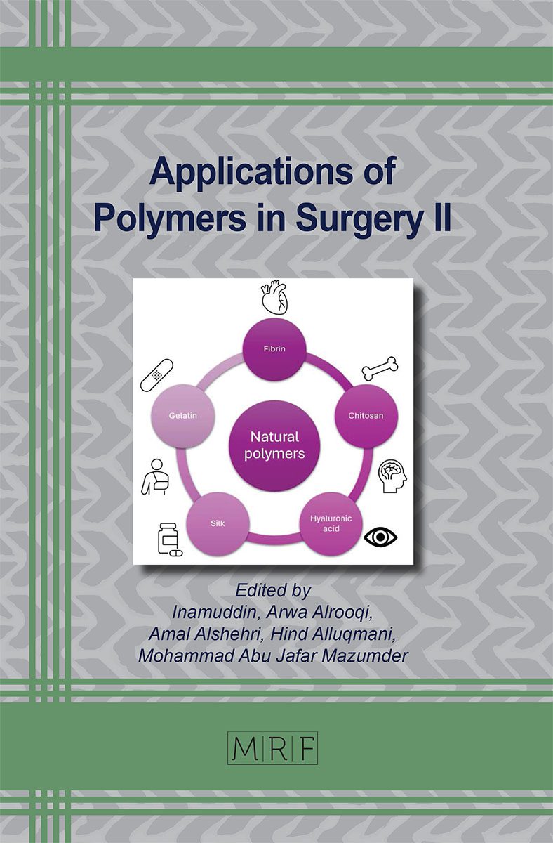
Part of the book on Applications of Polymers in Surgery II
References
[1] E. Kroll, D. Artzi, Enhancing aerospace engineering students’ learning with 3D printing wind‐tunnel models, Rapid Prototyp. J. 17 (2011) 393-402. https://doi.org/10.1108/13552541111156522
[2] P.F. Egan, Integrated design approaches for 3D printed tissue scaffolds: Review and outlook, Materials (Basel). 12 (2019) 2355. https://doi.org/10.3390/ma12152355
[3] T.D. Ngo, A. Kashani, G. Imbalzano, K.T.Q. Nguyen, D. Hui, Additive manufacturing (3D printing): A review of materials, methods, applications and challenges, Compos. Part B Eng. 143 (2018) 172-196. https://doi.org/10.1016/j.compositesb.2018.02.012
[4] C.L. Ventola, Medical applications for 3D printing: current and projected uses, Pharm. Ther. 39 (2014) 704.
[5] S.C. Ligon, R. Liska, J. Stampfl, M. Gurr, R. Mülhaupt, Polymers for 3D printing and customized additive manufacturing, Chem. Rev. 117 (2017) 10212-10290. https://doi.org/10.1021/acs.chemrev.7b00074
[6] P.F. Egan, K.A. Shea, S.J. Ferguson, Simulated tissue growth for 3D printed scaffolds, Biomech. Model. Mechanobiol. 17 (2018) 1481-1495. https://doi.org/10.1007/s10237-018-1040-9
[7] C. Y. Liaw, M. Guvendiren, Current and emerging applications of 3D printing in medicine, Biofabrication 9 (2017) 24102. https://doi.org/10.1088/1758-5090/aa7279
[8] J. Goole, K. Amighi, 3D printing in pharmaceutics: A new tool for designing customized drug delivery systems, Int. J. Pharm. 499 (2016) 376-394. https://doi.org/10.1016/j.ijpharm.2015.12.071
[9] I. Rubio-Pérez, A. Diaz Lantada, Surgical planning of sacral nerve stimulation procedure in presence of sacral anomalies by using personalized polymeric prototypes obtained with additive manufacturing techniques, Polymers (Basel). 12 (2020) 581. https://doi.org/10.3390/polym12030581
[10] J.M. Smit, A. Dimopoulou, A.G. Liss, C.J. Zeebregts, M. Kildal, I.S. Whitaker, A. Magnusson, R. Acosta, Preoperative CT angiography reduces surgery time in perforator flap reconstruction, J. Plast. Reconstr. Aesthetic Surg. 62 (2009) 1112-1117. https://doi.org/10.1016/j.bjps.2007.12.090
[11] W.M. Rozen, N.S. Anavekar, M.W. Ashton, D.L. Stella, D. Grinsell, R.J. Bloom, G.I. Taylor, Does the preoperative imaging of perforators with CT angiography improve operative outcomes in breast reconstruction?, Microsurg. Off. J. Int. Microsurg. Soc. Eur. Fed. Soc. Microsurg. 28 (2008) 516-523. https://doi.org/10.1002/micr.20526
[12] J. Masia, J.A. Clavero, J.R. Larranaga, X. Alomar, G. Pons, P. Serret, Multidetector-row computed tomography in the planning of abdominal perforator flaps, J. Plast. Reconstr. Aesthetic Surg. 59 (2006) 594-599. https://doi.org/10.1016/j.bjps.2005.10.024
[13] A. Alonso-Burgos, E. Garcia-Tutor, G. Bastarrika, D. Cano, A. Martinez-Cuesta, L. Pina, Preoperative planning of deep inferior epigastric artery perforator flap reconstruction with multislice-CT angiography: imaging findings and initial experience, J. Plast. Reconstr. Aesthetic Surg. 59 (2006) 585-593. https://doi.org/10.1016/j.bjps.2005.12.011
[14] J. Masia, D. Kosutic, D. Cervelli, J.A. Clavero, J.M. Monill, G. Pons, In search of the ideal method in perforator mapping: noncontrast magnetic resonance imaging, J. Reconstr. Microsurg. 26 (2010) 29-35. https://doi.org/10.1055/s-0029-1238222
[15] W.M. Rozen, D.L. Stella, J. Bowden, G.I. Taylor, M.W. Ashton, Advances in the pre‐operative planning of deep inferior epigastric artery perforator flaps: Magnetic resonance angiography, Microsurg. Off. J. Int. Microsurg. Soc. Eur. Fed. Soc. Microsurg. 29 (2009) 119-123. https://doi.org/10.1002/micr.20590
[16] K. V Wong, A. Hernandez, A review of additive manufacturing, Int. Sch. Res. Not. 2012 (2012) 208760. https://doi.org/10.5402/2012/208760
[17] E. Piskin, Biodegradable polymers as biomaterials, J. Biomater. Sci. Polym. Ed. 6 (1995) 775-795. https://doi.org/10.1163/156856295X00175
[18] R. Barbucci, Integrated biomaterials science, Springer Science & Business Media, 2002. https://doi.org/10.1007/b112196
[19] J.P. Vacanti, R. Langer, Tissue engineering: the design and fabrication of living replacement devices for surgical reconstruction and transplantation, Lancet 354 (1999) S32-S34. https://doi.org/10.1016/S0140-6736(99)90247-7
[20] T. Dvir, B.P. Timko, D.S. Kohane, R. Langer, Nanotechnological strategies for engineering complex tissues, Nano-Enabled Med. Appl. (2020) 351-382. https://doi.org/10.1201/9780429399039-12
[21] D.S. Kohane, R. Langer, Polymeric biomaterials in tissue engineering, Pediatr. Res. 63 (2008) 487-491. https://doi.org/10.1203/01.pdr.0000305937.26105.e7
[22] M. Vert, Aliphatic polyesters: great degradable polymers that cannot do everything, Biomacromolecules 6 (2005) 538-546. https://doi.org/10.1021/bm0494702
[23] S. Ramakrishna, J. Mayer, E. Wintermantel, K.W. Leong, Biomedical applications of polymer-composite materials: a review, Compos. Sci. Technol. 61 (2001) 1189-1224. https://doi.org/10.1016/S0266-3538(00)00241-4
[24] E.A. Guzzi, M.W. Tibbitt, Additive manufacturing of precision biomaterials, Adv. Mater. 32 (2020) 1901994. https://doi.org/10.1002/adma.201901994
[25] M. Colaco, D.A. Igel, A. Atala, The potential of 3D printing in urological research and patient care, Nat. Rev. Urol. 15 (2018) 213-221. https://doi.org/10.1038/nrurol.2018.6
[26] F. Rengier, A. Mehndiratta, H. Von Tengg-Kobligk, C.M. Zechmann, R. Unterhinninghofen, H.-U. Kauczor, F.L. Giesel, 3D printing based on imaging data: review of medical applications, Int. J. Comput. Assist. Radiol. Surg. 5 (2010) 335-341. https://doi.org/10.1007/s11548-010-0476-x
[27] K. Townsend, T. Pietila, 3D printing and modeling of congenital heart defects: a technical review, Birth Defects Res. 110 (2018) 1091-1097. https://doi.org/10.1002/bdr2.1342
[28] G. Lales, E. Anestiadou, V. Bisbinas, J.S. Suri, G. Tsoulfas, 3D printing: shedding light into the surgical education, in: 3D Print. Appl. Med. Surg., Elsevier, 2020: pp. 21-50. https://doi.org/10.1016/B978-0-323-66164-5.00003-9
[29] E.R. Perica, Z. Sun, A systematic review of three-dimensional printing in liver disease, J. Digit. Imaging 31 (2018) 692-701. https://doi.org/10.1007/s10278-018-0067-x
[30] J.S. Witowski, J. Coles-Black, T.Z. Zuzak, M. Pędziwiatr, J. Chuen, P. Major, A. Budzyński, 3D printing in liver surgery: a systematic review, Telemed. e-Health 23 (2017) 943-947. https://doi.org/10.1089/tmj.2017.0049
[31] Y. Nishihara, Y. Isobe, Y. Kitagawa, Validation of newly developed physical laparoscopy simulator in transabdominal preperitoneal (TAPP) inguinal hernia repair, Surg. Endosc. 31 (2017) 5429-5435. https://doi.org/10.1007/s00464-017-5614-x
[32] Y. Zheng, D. Yu, J. Zhao, Y. Wu, B. Zheng, 3D printout models vs. 3D-rendered images: which is better for preoperative planning?, J. Surg. Educ. 73 (2016) 518-523. https://doi.org/10.1016/j.jsurg.2016.01.003
[33] O.C. Burdall, E. Makin, M. Davenport, N. Ade-Ajayi, 3D printing to simulate laparoscopic choledochal surgery, J. Pediatr. Surg. 51 (2016) 828-831. https://doi.org/10.1016/j.jpedsurg.2016.02.093
[34] L. Kiraly, M. Tofeig, N.K. Jha, H. Talo, Three-dimensional printed prototypes refine the anatomy of post-modified Norwood-1 complex aortic arch obstruction and allow presurgical simulation of the repair, Interact. Cardiovasc. Thorac. Surg. 22 (2016) 238-240. https://doi.org/10.1093/icvts/ivv320
[35] I. Valverde, G. Gomez, J.F. Coserria, C. Suarez‐Mejias, S. Uribe, J. Sotelo, M.N. Velasco, J. Santos De Soto, A. Hosseinpour, T. Gomez‐Cia, 3 D printed models for planning endovascular stenting in transverse aortic arch hypoplasia, Catheter. Cardiovasc. Interv. 85 (2015) 1006-1012. https://doi.org/10.1002/ccd.25810
[36] K.A. Barsness, D.M. Rooney, L.M. Davis, E. O’Brien, Evaluation of three sources of validity evidence for a synthetic thoracoscopic esophageal atresia/tracheoesophageal fistula repair simulator, J. Laparoendosc. Adv. Surg. Tech. 25 (2015) 599-604. https://doi.org/10.1089/lap.2014.0370
[37] T. Akiba, T. Nakada, T. Inagaki, Simulation of the fissureless technique for thoracoscopic segmentectomy using rapid prototyping, Ann. Thorac. Cardiovasc. Surg. 21 (2015) 84-86. https://doi.org/10.5761/atcs.nm.13-00322
[38] D. Schmauss, C. Schmitz, A.K. Bigdeli, S. Weber, N. Gerber, A. Beiras-Fernandez, F. Schwarz, C. Becker, C. Kupatt, R. Sodian, Three-dimensional printing of models for preoperative planning and simulation of transcatheter valve replacement, Ann. Thorac. Surg. 93 (2012) e31-e33. https://doi.org/10.1016/j.athoracsur.2011.09.031
[39] T. Miyazaki, N. Yamasaki, T. Tsuchiya, K. Matsumoto, K. Takagi, T. Nagayasu, Airway stent insertion simulated with a three-dimensional printed airway model, Ann. Thorac. Surg. 99 (2015) e21-e23. https://doi.org/10.1016/j.athoracsur.2014.10.021
[40] K.A. Barsness, D.M. Rooney, L.M. Davis, E. O’Brien, Preliminary evaluation of a novel thoracoscopic infant lobectomy simulator, J. Laparoendosc. Adv. Surg. Tech. 25 (2015) 429-434. https://doi.org/10.1089/lap.2014.0364
[41] K. Takagi, A. Nanashima, T. Abo, J. Arai, N. Matsuo, T. Fukuda, T. Nagayasu, Three-dimensional printing model of liver for operative simulation in perihilar cholangiocarcinoma., Hepatogastroenterology. 61 (2014) 2315-2316.
[42] R. Souzaki, Y. Kinoshita, S. Ieiri, M. Hayashida, Y. Koga, K. Shirabe, T. Hara, Y. Maehara, M. Hashizume, T. Taguchi, Three-dimensional liver model based on preoperative CT images as a tool to assist in surgical planning for hepatoblastoma in a child, Pediatr. Surg. Int. 31 (2015) 593-596. https://doi.org/10.1007/s00383-015-3709-9
[43] D. Hoang, D. Perrault, M. Stevanovic, A. Ghiassi, Surgical applications of three-dimensional printing: a review of the current literature & how to get started, Ann. Transl. Med. 4 (2016). https://doi.org/10.21037/atm.2016.12.18
[44] K. Namba, A. Higaki, N. Kaneko, T. Mashiko, S. Nemoto, E. Watanabe, Microcatheter shaping for intracranial aneurysm coiling using the 3-dimensional printing rapid prototyping technology: preliminary result in the first 10 consecutive cases, World Neurosurg. 84 (2015) 178-186. https://doi.org/10.1016/j.wneu.2015.03.006
[45] M.D.B.S. Tam, S.D. Laycock, J.R.I. Brown, M. Jakeways, 3D printing of an aortic aneurysm to facilitate decision making and device selection for endovascular aneurysm repair in complex neck anatomy, J. Endovasc. Ther. 20 (2013) 863-867. https://doi.org/10.1583/13-4450MR.1
[46] K.J. Chung, D.Y. Hong, Y.T. Kim, I. Yang, Y.W. Park, H.N. Kim, Preshaping plates for minimally invasive fixation of calcaneal fractures using a real-size 3D-printed model as a preoperative and intraoperative tool, Foot Ankle Int. 35 (2014) 1231-1236. https://doi.org/10.1177/1071100714544522
[47] H.-S. Jeong, K.-J. Park, K.-M. Kil, S. Chong, H.-J. Eun, T.-S. Lee, J.-P. Lee, Minimally invasive plate osteosynthesis using 3D printing for shaft fractures of clavicles, Arch. Orthop. Trauma Surg. 134 (2014) 1551-1555. https://doi.org/10.1007/s00402-014-2075-8
[48] J.P. Costello, L.J. Olivieri, A. Krieger, O. Thabit, M.B. Marshall, S.-J. Yoo, P.C. Kim, R.A. Jonas, D.S. Nath, Utilizing three-dimensional printing technology to assess the feasibility of high-fidelity synthetic ventricular septal defect models for simulation in medical education, World J. Pediatr. Congenit. Hear. Surg. 5 (2014) 421-426. https://doi.org/10.1177/2150135114528721
[49] J. Soleman, F. Thieringer, J. Beinemann, C. Kunz, R. Guzman, Computer-assisted virtual planning and surgical template fabrication for frontoorbital advancement, Neurosurg. Focus 38 (2015) E5. https://doi.org/10.3171/2015.3.FOCUS14852
[50] W.Y. Angela, M. Khan, On-demand three-dimensional printing of surgical supplies in conflict zones, J. Trauma Acute Care Surg. 78 (2015) 201-203. https://doi.org/10.1097/TA.0000000000000481
[51] T.M. Rankin, N.A. Giovinco, D.J. Cucher, G. Watts, B. Hurwitz, D.G. Armstrong, Three-dimensional printing surgical instruments: are we there yet?, J. Surg. Res. 189 (2014) 193-197. https://doi.org/10.1016/j.jss.2014.02.020
[52] M. Maeda, N. Kanai, S. Kobayashi, T. Hosoi, R. Takagi, T. Ohki, Y. Muragaki, M. Yamato, S. Eguchi, F. Fukai, Endoscopic cell sheet transplantation device developed by using a 3-dimensional printer and its feasibility evaluation in a porcine model, Gastrointest. Endosc. 82 (2015) 147-152. https://doi.org/10.1016/j.gie.2015.01.062
[53] S. Kaneyama, T. Sugawara, M. Sumi, Safe and accurate midcervical pedicle screw insertion procedure with the patient-specific screw guide template system, Spine (Phila. Pa. 1976). 40 (2015) E341-E348. https://doi.org/10.1097/BRS.0000000000000772
[54] H. Chen, D. Wu, H. Yang, K. Guo, Clinical use of 3D printing guide plate in posterior lumbar pedicle screw fixation, Med. Sci. Monit. Int. Med. J. Exp. Clin. Res. 21 (2015) 3948. https://doi.org/10.12659/MSM.895597
[55] L. Ma, Y. Zhou, Y. Zhu, Z. Lin, Y. Wang, Y. Zhang, H. Xia, C. Mao, 3D-printed guiding templates for improved osteosarcoma resection, Sci. Rep. 6 (2016) 23335. https://doi.org/10.1038/srep23335
[56] M. Hirao, S. Ikemoto, H. Tsuboi, S. Akita, S. Ohshima, Y. Saeki, H. Yoshikawa, K. Sugamoto, T. Murase, J. Hashimoto, Computer assisted planning and custom-made surgical guide for malunited pronation deformity after first metatarsophalangeal joint arthrodesis in rheumatoid arthritis: a case report, Comput. Aided Surg. 19 (2014) 13-19. https://doi.org/10.3109/10929088.2014.885992
[57] Z. Jaffry, M. Masjedi, S. Clarke, S. Harris, M. Karia, B. Andrews, J. Cobb, Unicompartmental knee arthroplasties: robot vs. patient specific instrumentation, Knee 21 (2014) 428-434. https://doi.org/10.1016/j.knee.2013.11.017
[58] M. del Junco, Z. Okhunov, R. Yoon, R. Khanipour, S. Juncal, G. Abedi, A. Lusch, J. Landman, Development and initial porcine and cadaver experience with three-dimensional printing of endoscopic and laparoscopic equipment, J. Endourol. 29 (2015) 58-62. https://doi.org/10.1089/end.2014.0280
[59] A. Priester, S. Natarajan, J.D. Le, J. Garritano, B. Radosavcev, W. Grundfest, D.J.A. Margolis, L.S. Marks, J. Huang, A system for evaluating magnetic resonance imaging of prostate cancer using patient-specific 3D printed molds, Am. J. Clin. Exp. Urol. 2 (2014) 127.
[60] A. Shqaidef, A.F. Ayoub, B.S. Khambay, How accurate are rapid prototyped (RP) final orthognathic surgical wafers? A pilot study, Br. J. Oral Maxillofac. Surg. 52 (2014) 609-614. https://doi.org/10.1016/j.bjoms.2014.04.010
[61] S.-H. Kang, M.-K. Kim, B.C. Kim, S.-H. Lee, Orthognathic Y-splint: a CAD/CAM-engineered maxillary repositioning wafer assembly, Br. J. Oral Maxillofac. Surg. 52 (2014) 667-669. https://doi.org/10.1016/j.bjoms.2014.01.023
[62] N. Adolphs, W. Liu, E. Keeve, B. Hoffmeister, RapidSplint: virtual splint generation for orthognathic surgery-results of a pilot series, Comput. Aided Surg. 19 (2014) 20-28. https://doi.org/10.3109/10929088.2014.887778
[63] B. Li, L. Zhang, H. Sun, J. Yuan, S.G.F. Shen, X. Wang, A novel method of computer aided orthognathic surgery using individual CAD/CAM templates: a combination of osteotomy and repositioning guides, Br. J. Oral Maxillofac. Surg. 51 (2013) e239-e244. https://doi.org/10.1016/j.bjoms.2013.03.007
[64] M. Vehmeijer, M. van Eijnatten, N. Liberton, J. Wolff, A novel method of orbital floor reconstruction using virtual planning, 3-dimensional printing, and autologous bone, J. Oral Maxillofac. Surg. 74 (2016) 1608-1612. https://doi.org/10.1016/j.joms.2016.03.044
[65] A. Darwood, J. Collier, N. Joshi, W.E. Grant, V. Sauret-Jackson, R. Richards, A. Dawood, N. Kirkpatrick, Re-thinking 3D printing: a novel approach to guided facial contouring, J. Cranio-Maxillofacial Surg. 43 (2015) 1256-1260. https://doi.org/10.1016/j.jcms.2015.06.001
[66] A. Ruzza, M. Parekh, S. Ferrari, G. Salvalaio, Y. Nahum, C. Bovone, D. Ponzin, M. Busin, Preloaded donor corneal lenticules in a new validated 3D printed smart storage glide for Descemet stripping automated endothelial keratoplasty, Br. J. Ophthalmol. 99 (2015) 1388-1395. https://doi.org/10.1136/bjophthalmol-2014-306510
[67] M.L. Stitely, H. Paterson, Using three-dimensional printing to fabricate a tubing connector for dilation and evacuation, Obstet. Gynecol. 127 (2016) 317-319. https://doi.org/10.1097/AOG.0000000000001237
[68] A.B. AlAli, M.F. Griffin, P.E. Butler, Three-dimensional printing surgical applications, Eplasty 15 (2015).
[69] J. Li, M. Chen, X. Fan, H. Zhou, Recent advances in bioprinting techniques: approaches, applications and future prospects, J. Transl. Med. 14 (2016) 1-15. https://doi.org/10.1186/s12967-016-1028-0
[70] T. Goffin, P. Borry, K. Dierickx, H. Nys, Why eight EU Member States signed, but not yet ratified the Convention for Human Rights and Biomedicine, Health Policy (New. York). 86 (2008) 222-233. https://doi.org/10.1016/j.healthpol.2007.10.011
[71] L.K. Prasad, H. Smyth, 3D Printing technologies for drug delivery: a review, Drug Dev. Ind. Pharm. 42 (2016) 1019-1031. https://doi.org/10.3109/03639045.2015.1120743
[72] D. Kanters, A. de Vries, H. Boon, J. Urbach, A. Becht, H.-A. Kooistra, Quality assurance in medical 3D-printing, in: World Congr. Med. Phys. Biomed. Eng. 2018 June 3-8, 2018, Prague, Czech Repub. (Vol. 1), Springer, 2019: pp. 669-674. https://doi.org/10.1007/978-981-10-9035-6_125
[73] L. Adalbert, S.P.Y. Kanti, O. Jójárt-Laczkovich, H. Akel, I. Csóka, Expanding quality by design principles to support 3D printed medical device development following the renewed regulatory framework in Europe, Biomedicines 10 (2022) 2947. https://doi.org/10.3390/biomedicines10112947
[74] M. Kritikos, 3D Bio-Printing for Medical and Enhancement Purposes: Legal and Ethical Aspects, European Parliamentary Research Service (EPRS), 2018, (2018).
[75] F.L. Lopez, T.B. Ernest, C. Tuleu, M.O. Gul, Formulation approaches to pediatric oral drug delivery: benefits and limitations of current platforms, Expert Opin. Drug Deliv. 12 (2015) 1727-1740. https://doi.org/10.1517/17425247.2015.1060218
[76] S. Palekar, P.K. Nukala, S.M. Mishra, T. Kipping, K. Patel, Application of 3D printing technology and quality by design approach for development of age-appropriate pediatric formulation of baclofen, Int. J. Pharm. 556 (2019) 106-116. https://doi.org/10.1016/j.ijpharm.2018.11.062
[77] T. Persons, 3D Printing: Opportunities, Challenges, and Policy Implications of Additive Manufacturing, in: GAO-15-505Sp-Addictive Manuf. Forum, n. June, 2015: p. 63.
[78] C. Weller, R. Kleer, F.T. Piller, Economic implications of 3D printing: Market structure models in light of additive manufacturing revisited, Int. J. Prod. Econ. 164 (2015) 43-56. https://doi.org/10.1016/j.ijpe.2015.02.020
[79] J. Manners-Bell, K. Lyon, The implications of 3D printing for the global logistics industry, Transp. Intell. 1 (2012) 1-5.
[80] K. Cottrill, Transforming the future of supply chains through disruptive innovation, MIT Cent. Transp. Logist. Work. Pap. Spring (2011).
[81] M. Ye, The impact of 3D printing on the world container transport, (2015).
[82] T. Bartel, A. Rivard, A. Jimenez, C.A. Mestres, S. Müller, Medical three-dimensional printing opens up new opportunities in cardiology and cardiac surgery, Eur. Heart J. 39 (2018) 1246-1254. https://doi.org/10.1093/eurheartj/ehx016
[83] I.A. Ziogas, N.N. Zein, C. Quintini, C.M. Miller, G. Tsoulfas, Three-dimensional (3D) printing and liver transplantation, in: 3D Print. Appl. Med. Surg., Elsevier, 2020: pp. 97-116. https://doi.org/10.1016/B978-0-323-66164-5.00007-6
[84] D.A.B. Oliveira, R.Q. Feitosa, M.M. Correia, Segmentation of liver, its vessels and lesions from CT images for surgical planning, Biomed. Eng. Online 10 (2011) 1-23. https://doi.org/10.1186/1475-925X-10-30
[85] S.G. Farid, K.R. Prasad, G. Morris-Stiff, Operative terminology and post-operative management approaches applied to hepatic surgery: Trainee perspectives, World J. Gastrointest. Surg. 5 (2013) 146. https://doi.org/10.4240/wjgs.v5.i5.146
[86] K. Németh, R. Deshpande, Z. Máthé, A. Szuák, M. Kiss, C. Korom, Á. Nemeskéri, L. Kóbori, Extrahepatic arteries of the human liver-anatomical variants and surgical relevancies, Transpl. Int. 28 (2015) 1216-1226. https://doi.org/10.1111/tri.12630
[87] M. Uršič, M. Vrecl, G. Fazarinc, Corrosion cast study of the canine hepatic veins, Folia Morphol. (Warsz). 73 (2014) 475-481. https://doi.org/10.5603/FM.2014.0071
[88] M. Uršič, D. Ravnik, M. Hribernik, J. Pečar, J. Butinar, G. Fazarinc, Gross anatomy of the portal vein and hepatic artery ramifications in dogs: corrosion cast study, Anat. Histol. Embryol. 36 (2007) 83-87. https://doi.org/10.1111/j.1439-0264.2006.00719.x
[89] K.M. Farooqi, P.P. Sengupta, Echocardiography and three-dimensional printing: sound ideas to touch a heart, J. Am. Soc. Echocardiogr. 28 (2015) 398-403. https://doi.org/10.1016/j.echo.2015.02.005
[90] A. Shafiee, A. Atala, Printing technologies for medical applications, Trends Mol. Med. 22 (2016) 254-265. https://doi.org/10.1016/j.molmed.2016.01.003
[91] B. Mosadegh, G. Xiong, S. Dunham, J.K. Min, Current progress in 3D printing for cardiovascular tissue engineering, Biomed. Mater. 10 (2015) 34002. https://doi.org/10.1088/1748-6041/10/3/034002
[92] A.A. Giannopoulos, M.L. Steigner, E. George, M. Barile, A.R. Hunsaker, F.J. Rybicki, D. Mitsouras, Cardiothoracic applications of 3-dimensional printing, J. Thorac. Imaging 31 (2016) 253-272. https://doi.org/10.1097/RTI.0000000000000217
[93] R.K. Kankala, K. Zhu, J. Li, C.-S. Wang, S.-B. Wang, A.-Z. Chen, Fabrication of arbitrary 3D components in cardiac surgery: from macro-, micro-to nanoscale, Biofabrication 9 (2017) 32002. https://doi.org/10.1088/1758-5090/aa8113
[94] D. Shi, K. Liu, X. Zhang, H. Liao, X. Chen, Applications of three-dimensional printing technology in the cardiovascular field, Intern. Emerg. Med. 10 (2015) 769-780. https://doi.org/10.1007/s11739-015-1282-9
[95] S. Singare, Y. Liu, D. Li, B. Lu, S. He, Individually prefabricated prosthesis for maxilla reconstruction, J. Prosthodont. 17 (2008) 135-140. https://doi.org/10.1111/j.1532-849X.2007.00266.x
[96] S. Jacobs, R. Grunert, F.W. Mohr, V. Falk, 3D-Imaging of cardiac structures using 3D heart models for planning in heart surgery: a preliminary study, Interact. Cardiovasc. Thorac. Surg. 7 (2008) 6-9. https://doi.org/10.1510/icvts.2007.156588
[97] K. Subburaj, C. Nair, S. Rajesh, S.M. Meshram, B. Ravi, Rapid development of auricular prosthesis using CAD and rapid prototyping technologies, Int. J. Oral Maxillofac. Surg. 36 (2007) 938-943. https://doi.org/10.1016/j.ijom.2007.07.013
[98] R.F. Youssef, K. Spradling, R. Yoon, B. Dolan, J. Chamberlin, Z. Okhunov, R. Clayman, J. Landman, Applications of three‐dimensional printing technology in urological practice, BJU Int. 116 (2015) 697-702. https://doi.org/10.1111/bju.13183
[99] P. Bangeas, G. Voulalas, K. Ktenidis, Rapid prototyping in aortic surgery, Interact. Cardiovasc. Thorac. Surg. 22 (2016) 513-514. https://doi.org/10.1093/icvts/ivv395
[100] D. Ho, A. Squelch, Z. Sun, Modelling of aortic aneurysm and aortic dissection through 3D printing, J. Med. Radiat. Sci. 64 (2017) 10-17. https://doi.org/10.1002/jmrs.212
[101] J.F.B. Chick, S.N. Reddy, C.Y. Alice, T. Kelil, R.N. Srinivasa, K.J. Cooper, W.E. Saad, Three-dimensional printing facilitates successful endovascular closure of a type II abernethy malformation using an Amplatzer Atrial Septal Occluder device, Ann. Vasc. Surg. 43 (2017) 311-e15. https://doi.org/10.1016/j.avsg.2017.02.012
[102] D. Maragiannis, M.S. Jackson, S.R. Igo, R.C. Schutt, P. Connell, J. Grande-Allen, C.M. Barker, S.M. Chang, M.J. Reardon, W.A. Zoghbi, Replicating patient-specific severe aortic valve stenosis with functional 3D modeling, Circ. Cardiovasc. Imaging 8 (2015) e003626. https://doi.org/10.1161/CIRCIMAGING.115.003626
[103] D. Maragiannis, M.S. Jackson, S.R. Igo, S.M. Chang, W.A. Zoghbi, S.H. Little, Functional 3D printed patient-specific modeling of severe aortic stenosis, J. Am. Coll. Cardiol. 64 (2014) 1066-1068. https://doi.org/10.1016/j.jacc.2014.05.058
[104] S. Schievano, F. Migliavacca, L. Coats, S. Khambadkone, M. Carminati, N. Wilson, J.E. Deanfield, P. Bonhoeffer, A.M. Taylor, Percutaneous pulmonary valve implantation based on rapid prototyping of right ventricular outflow tract and pulmonary trunk from MR data, Radiology 242 (2007) 490-497. https://doi.org/10.1148/radiol.2422051994
[105] S. Milosavljevic, P.D. Milburn, B.W. Knox, The influence of occupation on lumbar sagittal motion and posture, Ergonomics 48 (2005) 657-667. https://doi.org/10.1080/00140130500070848
[106] H. Takao, S. Amemiya, E. Shibata, K. Ohtomo, Three-Dimensional Printing of Hollow Portal Vein Stenosis Models: A Feasibility Study., J. Vasc. Interv. Radiol. JVIR 27 (2016) 1755-1758. https://doi.org/10.1016/j.jvir.2016.05.022
[107] C. Chevallier, W. Willaert, E. Kawa, M. Centola, B. Steger, R. Dirnhofer, P. Mangin, S. Grabherr, Postmortem circulation: a new model for testing endovascular devices and training clinicians in their use, Clin. Anat. 27 (2014) 556-562. https://doi.org/10.1002/ca.22357
[108] X. Han, R. Bibb, R. Harris, Engineering design of artificial vascular junctions for 3D printing, Biofabrication 8 (2016) 25018. https://doi.org/10.1088/1758-5090/8/2/025018
[109] K. Sommer, R.L. Izzo, L. Shepard, A.R. Podgorsak, S. Rudin, A.H. Siddiqui, M.F. Wilson, E. Angel, Z. Said, M. Springer, Design optimization for accurate flow simulations in 3D printed vascular phantoms derived from computed tomography angiography, in: Med. Imaging 2017 Imaging Informatics Heal. Res. Appl., SPIE, 2017: pp. 180-191. https://doi.org/10.1117/12.2253711
[110] C. Canstein, P. Cachot, A. Faust, A.F. Stalder, J. Bock, A. Frydrychowicz, J. Küffer, J. Hennig, M. Markl, 3D MR flow analysis in realistic rapid‐prototyping model systems of the thoracic aorta: comparison with in vivo data and computational fluid dynamics in identical vessel geometries, Magn. Reson. Med. An Off. J. Int. Soc. Magn. Reson. Med. 59 (2008) 535-546. https://doi.org/10.1002/mrm.21331
[111] R.R. Ahmadian, A.P. Boyd, A.M. Scotti, J.D. Collins, J.C. Carr, P.M. McCarthy, S.C. Malaisrie, A.J. Barker, M. Markl, Comprehensive evaluation of aortic disease by in-vivo 4D flow MRI and 3D printing of patient-specific models: a feasibility study, J. Cardiovasc. Magn. Reson. 18 (2016) 1-3. https://doi.org/10.1186/s12968-015-0221-2
[112] N. Martelli, C. Serrano, H. van den Brink, J. Pineau, P. Prognon, I. Borget, S. El Batti, Advantages and disadvantages of 3-dimensional printing in surgery: a systematic review, Surgery 159 (2016) 1485-1500. https://doi.org/10.1016/j.surg.2015.12.017
[113] R. Vaishya, M.K. Patralekh, A. Vaish, A.K. Agarwal, V. Vijay, Publication trends and knowledge mapping in 3D printing in orthopaedics, J. Clin. Orthop. Trauma 9 (2018) 194-201. https://doi.org/10.1016/j.jcot.2018.07.006
[114] V. Bagaria, S. Deshpande, D.D. Rasalkar, A. Kuthe, B.K. Paunipagar, Use of rapid prototyping and three-dimensional reconstruction modeling in the management of complex fractures, Eur. J. Radiol. 80 (2011) 814-820. https://doi.org/10.1016/j.ejrad.2010.10.007
[115] C. Hurson, A. Tansey, B. O’donnchadha, P. Nicholson, J. Rice, J. McElwain, Rapid prototyping in the assessment, classification and preoperative planning of acetabular fractures, Injury 38 (2007) 1158-1162. https://doi.org/10.1016/j.injury.2007.05.020
[116] H. Lal, M.K. Patralekh, 3D printing and its applications in orthopaedic trauma: a technological marvel, J. Clin. Orthop. Trauma 9 (2018) 260-268. https://doi.org/10.1016/j.jcot.2018.07.022
[117] T.M. Wong, J. Jin, T.W. Lau, C. Fang, C.H. Yan, K. Yeung, M. To, F. Leung, The use of three-dimensional printing technology in orthopaedic surgery: a review, J. Orthop. Surg. 25 (2017) 2309499016684077. https://doi.org/10.1177/2309499016684077
[118] L. Wan, X. Zhang, S. Zhang, K. Li, P. Cao, J. Li, G. Wu, Clinical feasibility and application value of computer virtual reduction combined with 3D printing technique in complex acetabular fractures, Exp. Ther. Med. 17 (2019) 3630-3636. https://doi.org/10.3892/etm.2019.7344
[119] Z. Liu, J. Jia, Y. Zhang, W. Tian, X. Jin, Y. Hu, Internal fixation of complicated acetabular fractures directed by preoperative surgery with 3D printing models, Orthop. Surg. 9 (2017) 257-260. https://doi.org/10.1111/os.12324
[120] H.J. Kang, B.S. Kim, S.M. Kim, Y.M. Kim, H.N. Kim, J.Y. Park, J.H. Cho, Y. Choi, Can preoperative 3D printing change surgeon’s operative plan for distal tibia fracture?, Biomed Res. Int. 2019 (2019) 7059413. https://doi.org/10.1155/2019/7059413
[121] C.M. Wixted, J.R. Peterson, R.J. Kadakia, S.B. Adams, Three-dimensional printing in orthopaedic surgery: current applications and future developments, JAAOS Glob. Res. Rev. 5 (2021) e20. https://doi.org/10.5435/JAAOSGlobal-D-20-00230
[122] B. Garg, M. Gupta, M. Singh, D. Kalyanasundaram, Outcome and safety analysis of 3D-printed patient-specific pedicle screw jigs for complex spinal deformities: a comparative study, Spine J. 19 (2019) 56-64. https://doi.org/10.1016/j.spinee.2018.05.001
[123] W. Cho, A.V. Job, J. Chen, J.H. Baek, A review of current clinical applications of three-dimensional printing in spine surgery, Asian Spine J. 12 (2018) 171. https://doi.org/10.4184/asj.2018.12.1.171
[124] J.S. Mulford, S. Babazadeh, N. Mackay, Three‐dimensional printing in orthopaedic surgery: review of current and future applications, ANZ J. Surg. 86 (2016) 648-653. https://doi.org/10.1111/ans.13533
[125] K.C. Wong, 3D-printed patient-specific applications in orthopedics, Orthop. Res. Rev. (2016) 57-66. https://doi.org/10.2147/ORR.S99614
[126] A.E.M. Eltorai, E. Nguyen, A.H. Daniels, Three-dimensional printing in orthopedic surgery, Orthopedics 38 (2015) 684-687. https://doi.org/10.3928/01477447-20151016-05
[127] A. Aimar, A. Palermo, B. Innocenti, The role of 3D printing in medical applications: a state of the art, J. Healthc. Eng. 2019 (2019) 5340616. https://doi.org/10.1155/2019/5340616
[128] C.L. Jeng, J.T. Campbell, E.Y. Tang, R.A. Cerrato, M.S. Myerson, Tibiotalocalcaneal arthrodesis with bulk femoral head allograft for salvage of large defects in the ankle, Foot Ankle Int. 34 (2013) 1256-1266. https://doi.org/10.1177/1071100713488765
[129] P. Canzi, M. Magnetto, S. Marconi, P. Morbini, S. Mauramati, F. Aprile, I. Avato, F. Auricchio, M. Benazzo, New frontiers and emerging applications of 3D printing in ENT surgery: a systematic review of the literature, Acta Otorhinolaryngol. Ital. 38 (2018) 286. https://doi.org/10.14639/0392-100X-1984
[130] P.A. Yushkevich, J. Piven, H.C. Hazlett, R.G. Smith, S. Ho, J.C. Gee, G. Gerig, User-guided 3D active contour segmentation of anatomical structures: significantly improved efficiency and reliability, Neuroimage 31 (2006) 1116-1128. https://doi.org/10.1016/j.neuroimage.2006.01.015
[131] A.S. Rose, C.E. Webster, O.L.A. Harrysson, E.J. Formeister, R.B. Rawal, C.E. Iseli, Pre-operative simulation of pediatric mastoid surgery with 3D-printed temporal bone models, Int. J. Pediatr. Otorhinolaryngol. 79 (2015) 740-744. https://doi.org/10.1016/j.ijporl.2015.03.004
[132] S.E. Mowry, H. Jammal, C. Myer IV, C.A. Solares, P. Weinberger, A novel temporal bone simulation model using 3D printing techniques, Otol. Neurotol. 36 (2015) 1562-1565. https://doi.org/10.1097/MAO.0000000000000848
[133] J.B. Hochman, C. Rhodes, D. Wong, J. Kraut, J. Pisa, B. Unger, Comparison of cadaveric and isomorphic three‐dimensional printed models in temporal bone education, Laryngoscope 125 (2015) 2353-2357. https://doi.org/10.1002/lary.24919
[134] M.J. Da Cruz, H.W. Francis, Face and content validation of a novel three-dimensional printed temporal bone for surgical skills development, J. Laryngol. Otol. 129 (2015) S23-S29. https://doi.org/10.1017/S0022215115001346
[135] J.B. Hochman, J. Kraut, K. Kazmerik, B.J. Unger, Generation of a 3D printed temporal bone model with internal fidelity and validation of the mechanical construct, Otolaryngol. Neck Surg. 150 (2014) 448-454. https://doi.org/10.1177/0194599813518008
[136] C.M. Chiesa Estomba, I. González Fernández, M.Á. Iglesias Otero, How we do it: anterior and posterior nosebleed trainer, the 3D printing epistaxis project, Clin Otolaryngol 25 (2016) 1-2. https://doi.org/10.1111/coa.12711
[137] C.M. Johnson, J.T. Howell, D.J. Mettenburg, F.A. Rueggeberg, R.J. Howell, G.N. Postma, P.M. Weinberger, Mechanical modeling of the human cricoid cartilage using computer-aided design: applications in airway balloon dilation research, Ann. Otol. Rhinol. Laryngol. 125 (2016) 69-76. https://doi.org/10.1177/0003489415598999
[138] K.R. Kavanagh, V. Cote, Y. Tsui, S. Kudernatsch, D.R. Peterson, T.A. Valdez, Pediatric laryngeal simulator using 3D printed models: a novel technique, Laryngoscope 127 (2017) E132-E137. https://doi.org/10.1002/lary.26326
[139] S. Bustamante, S. Bose, P. Bishop, R. Klatte, F. Norris, Novel application of rapid prototyping for simulation of bronchoscopic anatomy, J. Cardiothorac. Vasc. Anesth. 28 (2014) 1122-1125. https://doi.org/10.1053/j.jvca.2013.08.015
[140] T.A. Ainsworth, J.B. Kobler, G.J. Loan, J.A. Burns, Simulation model for transcervical laryngeal injection providing real-time feedback, Ann. Otol. Rhinol. Laryngol. 123 (2014) 881-886. https://doi.org/10.1177/0003489414539922
[141] D. Rokaya, V. Srimaneepong, J. Sapkota, J. Qin, K. Siraleartmukul, V. Siriwongrungson, Polymeric materials and films in dentistry: An overview, J. Adv. Res. 14 (2018) 25-34. https://doi.org/10.1016/j.jare.2018.05.001
[142] H.-H. Lin, D. Lonic, L.-J. Lo, 3D printing in orthognathic surgery− A literature review, J. Formos. Med. Assoc. 117 (2018) 547-558. https://doi.org/10.1016/j.jfma.2018.01.008
[143] C.M. Cristache, S. Gurbanescu, Accuracy evaluation of a stereolithographic surgical template for dental implant insertion using 3D superimposition protocol, Int. J. Dent. 2017 (2017) 4292081. https://doi.org/10.1155/2017/4292081
[144] A. Tahmaseb, D. Wismeijer, W. Coucke, W. Derksen, Computer technology applications in surgical implant dentistry: a systematic review, Int J Oral Maxillofac Implant. 29 (2014) 25-42. https://doi.org/10.11607/jomi.2014suppl.g1.2
[145] D.A. Tyndall, J.B. Price, S. Tetradis, S.D. Ganz, C. Hildebolt, W.C. Scarfe, Position statement of the American Academy of Oral and Maxillofacial Radiology on selection criteria for the use of radiology in dental implantology with emphasis on cone beam computed tomography, Oral Surg. Oral Med. Oral Pathol. Oral Radiol. 113 (2012) 817-826. https://doi.org/10.1016/j.oooo.2012.03.005
[146] M. Klein, M. Abrams, Computer-guided surgery utilizing a computer-milled surgical template., Pract. Proced. Aesthetic Dent. PPAD 13 (2001) 165-169.
[147] M. Revilla-León, M. Sadeghpour, M. Özcan, An update on applications of 3D printing technologies used for processing polymers used in implant dentistry, Odontology 108 (2020) 331-338. https://doi.org/10.1007/s10266-019-00441-7
[148] P.B. Tardieu, L. Vrielinck, E. Escolano, Computer-assisted implant placement. A case report: treatment of the mandible., Int. J. Oral Maxillofac. Implants 18 (2003).
[149] G.A. Mandelaris, S.D. Vlk, Guided implant surgery with placement of a presurgical CAD/CAM patient-specific abutment and provisional in the esthetic zone, Compend Contin Educ Dent 35 (2014) 494-504.
[150] L. Tapie, N. Lebon, B. Mawussi, F. Duret, J.-P. Attal, Understanding dental CAD/CAM for restorations–the digital workflow from a mechanical engineering viewpoint., Int. J. Comput. Dent. 18 (2015) 21-44.
[151] S.D. Ganz, Three-dimensional imaging and guided surgery for dental implants, Dent. Clin. 59 (2015) 265-290. https://doi.org/10.1016/j.cden.2014.11.001
[152] M. Javaid, A. Haleem, 4D printing applications in medical field: a brief review, Clin. Epidemiol. Glob. Heal. 7 (2019) 317-321. https://doi.org/10.1016/j.cegh.2018.09.007
[153] C. Schubert, M.C. Van Langeveld, L.A. Donoso, Innovations in 3D printing: a 3D overview from optics to organs, Br. J. Ophthalmol. 98 (2014) 159-161. https://doi.org/10.1136/bjophthalmol-2013-304446
[154] G.E. Cacciamani, Z. Okhunov, A.D. Meneses, M.E. Rodriguez-Socarras, J.G. Rivas, F. Porpiglia, E. Liatsikos, D. Veneziano, Impact of three-dimensional printing in urology: state of the art and future perspectives. A systematic review by ESUT-YAUWP Group, Eur. Urol. 76 (2019) 209-221. https://doi.org/10.1016/j.eururo.2019.04.044
[155] E. Checcucci, D. Amparore, C. Fiori, M. Manfredi, M. Ivano, M. Di Dio, G. Niculescu, F. Piramide, G. Cattaneo, P. Piazzolla, 3D imaging applications for robotic urologic surgery: an ESUT YAUWP review, World J. Urol. 38 (2020) 869-881. https://doi.org/10.1007/s00345-019-02922-4
[156] R. Souzaki, Y. Kinoshita, S. Ieiri, N. Kawakubo, S. Obata, T. Jimbo, Y. Koga, M. Hashizume, T. Taguchi, Preoperative surgical simulation of laparoscopic adrenalectomy for neuroblastoma using a three-dimensional printed model based on preoperative CT images, J. Pediatr. Surg. 50 (2015) 2112-2115. https://doi.org/10.1016/j.jpedsurg.2015.08.037
[157] J.L. Silberstein, M.M. Maddox, P. Dorsey, A. Feibus, R. Thomas, B.R. Lee, Physical models of renal malignancies using standard cross-sectional imaging and 3-dimensional printers: a pilot study, Urology 84 (2014) 268-273. https://doi.org/10.1016/j.urology.2014.03.042
[158] M.E. Westerman, J.M. Matsumoto, J.M. Morris, B.C. Leibovich, Three-dimensional printing for renal cancer and surgical planning, Eur. Urol. Focus 2 (2016) 574-576. https://doi.org/10.1016/j.euf.2016.12.009
[159] P. Chandak, N. Byrne, H. Lynch, C. Allen, G. Rottenberg, A. Chandra, N. Raison, H. Ahmed, V. Kasivisvanathan, O. Elhage, Three-dimensional printing in robot-assisted radical prostatectomy-an Idea, Development, Exploration, Assessment, Long-term follow-up (IDEAL) Phase 2a study, BJU Int. 122 (2018) 360-361. https://doi.org/10.1111/bju.14189
[160] N. Wake, T. Rude, S.K. Kang, M.D. Stifelman, J.F. Borin, D.K. Sodickson, W.C. Huang, H. Chandarana, 3D printed renal cancer models derived from MRI data: application in pre-surgical planning, Abdom. Radiol. 42 (2017) 1501-1509. https://doi.org/10.1007/s00261-016-1022-2
[161] T. Smektala, A. Gołab, M. Królikowski, M. Słojewski, Low cost silicone renal replicas for surgical training. Technical note, Arch. Españoles Urol. 69 (2016) 434-436.
[162] A.E. Ghazi, B.A. Teplitz, Role of 3D printing in surgical education for robotic urology procedures, Transl. Androl. Urol. 9 (2020) 931. https://doi.org/10.21037/tau.2020.01.03
[163] M.C. Dewan, A. Rattani, S. Gupta, R.E. Baticulon, Y.-C. Hung, M. Punchak, A. Agrawal, A.O. Adeleye, M.G. Shrime, A.M. Rubiano, Estimating the global incidence of traumatic brain injury, J. Neurosurg. 130 (2018) 1080-1097. https://doi.org/10.3171/2017.10.JNS17352
[164] A.I.R. Maas, D.K. Menon, P.D. Adelson, N. Andelic, M.J. Bell, A. Belli, P. Bragge, A. Brazinova, A. Büki, R.M. Chesnut, Traumatic brain injury: integrated approaches to improve prevention, clinical care, and research, Lancet Neurol. 16 (2017) 987-1048.
[165] M.E. Ring, The history of maxillofacial prosthetics, Plast. Reconstr. Surg. 87 (1991) 174-184. https://doi.org/10.1097/00006534-199101000-00031
[166] R. Lanzara, M. Viswambaran, D. Kumar, Maxillofacial prosthetic materials: current status and recent advances: A comprehensive review, Int. J. Appl. Dent. Sci 7 (2021) 255-259. https://doi.org/10.22271/oral.2021.v7.i2d.1219
[167] C.H. Pritham, Fundamentals of facial prosthetics, JPO J. Prosthetics Orthot. 7 (1995) 22A. https://doi.org/10.1097/00008526-199507040-00011
[168] D. Palousek, J. Rosicky, D. Koutny, Use of digital technologies for nasal prosthesis manufacturing, Prosthet. Orthot. Int. 38 (2014) 171-175. https://doi.org/10.1177/0309364613489333
[169] E.J. Bos, T. Scholten, Y. Song, J.C. Verlinden, J. Wolff, T. Forouzanfar, M.N. Helder, P. Van Zuijlen, Developing a parametric ear model for auricular reconstruction: a new step towards patient-specific implants, J. Cranio-Maxillofacial Surg. 43 (2015) 390-395. https://doi.org/10.1016/j.jcms.2014.12.016
[170] Y.L. Yap, Y.S.E. Tan, H.K.J. Tan, Z.K. Peh, X.Y. Low, W.Y. Yeong, C.S.H. Tan, A. Laude, 3D printed bio-models for medical applications, Rapid Prototyp. J. 23 (2017) 227-235. https://doi.org/10.1108/RPJ-08-2015-0102
[171] C. Kincade, L. McHutchion, J. Wolfaardt, Digital design of patient-specific abutments for the retention of implant-retained facial prostheses, J. Prosthet. Dent. 120 (2018) 309-312. https://doi.org/10.1016/j.prosdent.2017.10.002
[172] P. Tack, J. Victor, P. Gemmel, L. Annemans, 3D-printing techniques in a medical setting: a systematic literature review, Biomed. Eng. Online 15 (2016) 1-21. https://doi.org/10.1186/s12938-015-0119-0
[173] E. Provaggi, J.J.H. Leong, D.M. Kalaskar, Applications of 3D printing in the management of severe spinal conditions, Proc. Inst. Mech. Eng. Part H J. Eng. Med. 231 (2017) 471-486. https://doi.org/10.1177/0954411916667761
[174] A.-M. Wu, Z.-X. Shao, J.-S. Wang, X.-D. Yang, W.-Q. Weng, X.-Y. Wang, H.-Z. Xu, Y.-L. Chi, Z.-K. Lin, The accuracy of a method for printing three-dimensional spinal models, PLoS One 10 (2015) e0124291. https://doi.org/10.1371/journal.pone.0124291
[175] V. Baskaran, G. Štrkalj, M. Štrkalj, A. Di Ieva, Current applications and future perspectives of the use of 3D printing in anatomical training and neurosurgery, Front. Neuroanat. 10 (2016) 69. https://doi.org/10.3389/fnana.2016.00069
[176] F. Guo, J. Dai, J. Zhang, Y. Ma, G. Zhu, J. Shen, G. Niu, Individualized 3D printing navigation template for pedicle screw fixation in upper cervical spine, PLoS One 12 (2017) e0171509. https://doi.org/10.1371/journal.pone.0171509
[177] Z. Wu, L. Huang, H. Sang, Z. Ma, S. Wan, G. Cui, W. Lei, Accuracy and safety assessment of pedicle screw placement using the rapid prototyping technique in severe congenital scoliosis, Clin. Spine Surg. 24 (2011) 444-450. https://doi.org/10.1097/BSD.0b013e318201be2a
[178] K. Mao, Y. Wang, S. Xiao, Z. Liu, Y. Zhang, X. Zhang, Z. Wang, N. Lu, Z. Shourong, Z. Xifeng, Clinical application of computer-designed polystyrene models in complex severe spinal deformities: a pilot study, Eur. Spine J. 19 (2010) 797-802. https://doi.org/10.1007/s00586-010-1359-0
[179] P.S. D’Urso, G. Askin, J.S. Earwaker, G.S. Merry, R.G. Thompson, T.M. Barker, D.J. Effeney, Spinal biomodeling, Spine (Phila. Pa. 1976). 24 (1999) 1247-1251. https://doi.org/10.1097/00007632-199906150-00013
[180] P.S. D’Urso, O.D. Williamson, R.G. Thompson, Biomodeling as an aid to spinal instrumentation, Spine (Phila. Pa. 1976). 30 (2005) 2841-2845. https://doi.org/10.1097/01.brs.0000190886.56895.3d
[181] Y.-T. Wang, X.-J. Yang, B. Yan, T.-H. Zeng, Y.-Y. Qiu, S.-J. Chen, Clinical application of three-dimensional printing in the personalized treatment of complex spinal disorders, Chinese J. Traumatol. 19 (2016) 31-34. https://doi.org/10.1016/j.cjtee.2015.09.009
[182] M. Yang, C. Li, Y. Li, Y. Zhao, X. Wei, G. Zhang, J. Fan, H. Ni, Z. Chen, Y. Bai, Application of 3D rapid prototyping technology in posterior corrective surgery for Lenke 1 adolescent idiopathic scoliosis patients, Medicine (Baltimore). 94 (2015) e582. https://doi.org/10.1097/MD.0000000000000582
[183] C. Li, M. Yang, Y. Xie, Z. Chen, C. Wang, Y. Bai, X. Zhu, M. Li, Application of the polystyrene model made by 3-D printing rapid prototyping technology for operation planning in revision lumbar discectomy, J. Orthop. Sci. 20 (2015) 475-480. https://doi.org/10.1007/s00776-015-0706-8
[184] K. Toyoda, E. Urasaki, Y. Yamakawa, Novel approach for the efficient use of a full-scale, 3-dimensional model for cervical posterior fixation: a technical case report, Spine (Phila. Pa. 1976). 38 (2013) E1357-E1360. https://doi.org/10.1097/BRS.0b013e3182a1f1bd
[185] M.T. Izatt, P.L.P.J. Thorpe, R.G. Thompson, P.S. D’Urso, C.J. Adam, J.W.S. Earwaker, R.D. Labrom, G.N. Askin, The use of physical biomodelling in complex spinal surgery, Eur. Spine J. 16 (2007) 1507-1518. https://doi.org/10.1007/s00586-006-0289-3
[186] J. Guarino, S. Tennyson, G. McCain, L. Bond, K. Shea, H. King, Rapid prototyping technology for surgeries of the pediatric spine and pelvis: benefits analysis, J. Pediatr. Orthop. 27 (2007) 955-960. https://doi.org/10.1097/bpo.0b013e3181594ced
[187] P.S. D’Urso, T.M. Barker, W.J. Earwaker, L.J. Bruce, R.L. Atkinson, M.W. Lanigan, J.F. Arvier, D.J. Effeney, Stereolithographic biomodelling in cranio-maxillofacial surgery: a prospective trial, J. Cranio-Maxillofacial Surg. 27 (1999) 30-37. https://doi.org/10.1016/S1010-5182(99)80007-9
[188] M.P. Kim, A.H. Ta, W.A. Ellsworth IV, R.A. Marco, P. Gaur, J.S. Miller, Three dimensional model for surgical planning in resection of thoracic tumors, Int. J. Surg. Case Rep. 16 (2015) 127-129. https://doi.org/10.1016/j.ijscr.2015.09.037
[189] A. Goel, B. Jankharia, A. Shah, P. Sathe, Three-dimensional models: an emerging investigational revolution for craniovertebral junction surgery, J. Neurosurg. Spine 25 (2016) 740-744. https://doi.org/10.3171/2016.4.SPINE151268
[190] J. Mizutani, T. Matsubara, M. Fukuoka, N. Tanaka, H. Iguchi, A. Furuya, H. Okamoto, I. Wada, T. Otsuka, Application of full-scale three-dimensional models in patients with rheumatoid cervical spine, Eur. Spine J. 17 (2008) 644-649. https://doi.org/10.1007/s00586-008-0611-3
[191] Y. Liew, E. Beveridge, A.K. Demetriades, M.A. Hughes, 3D printing of patient-specific anatomy: a tool to improve patient consent and enhance imaging interpretation by trainees, Br. J. Neurosurg. 29 (2015) 712-714. https://doi.org/10.3109/02688697.2015.1026799
[192] J.C. Yang, X.Y. Ma, J. Lin, Z.H. Wu, K. Zhang, Q.S. Yin, Personalised modified osteotomy using computer-aided design-rapid prototyping to correct thoracic deformities, Int. Orthop. 35 (2011) 1827-1832. https://doi.org/10.1007/s00264-010-1155-9
[193] I. Madrazo, C. Zamorano, E. Magallón, T. Valenzuela, A. Ibarra, H. Salgado-Ceballos, I. Grijalva, R.E. Franco-Bourland, G. Guízar-Sahagún, Stereolithography in spine pathology: a 2-case report, Surg. Neurol. 72 (2009) 272-275. https://doi.org/10.1016/j.surneu.2008.04.034
[194] P.A. Anderson, Clinical applications of 3D printing, Spine (Phila. Pa. 1976). 42 (2017) S30-S31. https://doi.org/10.1097/BRS.0000000000002039
[195] T. Sugawara, N. Higashiyama, S. Kaneyama, M. Sumi, Accurate and simple screw insertion procedure with patient-specific screw guide templates for posterior C1-C2 fixation, Spine (Phila. Pa. 1976). 42 (2017) E340-E346. https://doi.org/10.1097/BRS.0000000000001807
[196] T. Deng, M. Jiang, Q. Lei, L. Cai, L. Chen, The accuracy and the safety of individualized 3D printing screws insertion templates for cervical screw insertion, Comput. Assist. Surg. 21 (2016) 143-149. https://doi.org/10.1080/24699322.2016.1236146
[197] B.D. Owen, G.E. Christensen, J.M. Reinhardt, T.C. Ryken, Rapid prototype patient-specific drill template for cervical pedicle screw placement, Comput. Aided Surg. 12 (2007) 303-308. https://doi.org/10.3109/10929080701662826
[198] K. Liu, Q. Zhang, X. Li, C. Zhao, X. Quan, R. Zhao, Z. Chen, Y. Li, Preliminary application of a multi-level 3D printing drill guide template for pedicle screw placement in severe and rigid scoliosis, Eur. Spine J. 26 (2017) 1684-1689. https://doi.org/10.1007/s00586-016-4926-1
[199] S. Lu, Y.Q. Xu, Y.Z. Zhang, Y.B. Li, L.E. Xie, J.H. Shi, H. Guo, G.P. Chen, Y.B. Chen, A novel computer‐assisted drill guide template for lumbar pedicle screw placement: a cadaveric and clinical study, Int. J. Med. Robot. Comput. Assist. Surg. 5 (2009) 184-191. https://doi.org/10.1002/rcs.249
[200] A.-M. Wu, S. Wang, W.-Q. Weng, Z.-X. Shao, X.-D. Yang, J.-S. Wang, H.-Z. Xu, Y.-L. Chi, The radiological feature of anterior occiput-to-axis screw fixation as it guides the screw trajectory on 3D printed models: a feasibility study on 3D images and 3D printed models, Medicine (Baltimore). 93 (2014) e242. https://doi.org/10.1097/MD.0000000000000242
[201] T. Lu, C. Liu, J. Dong, M. Lu, H. Li, X. He, Cervical screw placement using rapid prototyping drill templates for navigation: a literature review, Int. J. Comput. Assist. Radiol. Surg. 11 (2016) 2231-2240. https://doi.org/10.1007/s11548-016-1414-3
[202] K.-Y. Huang, R.-M. Lin, J.-J. Fang, A novel method of C1-C2 transarticular screw insertion for symptomatic atlantoaxial instability using a customized guiding block: A case report and a technical note, Medicine (Baltimore). 95 (2016) e5100. https://doi.org/10.1097/MD.0000000000005100
[203] S. Lu, Y.Q. Xu, G.P. Chen, Y.Z. Zhang, D. Lu, Y.B. Chen, J.H. Shi, X.M. Xu, Efficacy and accuracy of a novel rapid prototyping drill template for cervical pedicle screw placement, Comput. Aided Surg. 16 (2011) 240-248. https://doi.org/10.3109/10929088.2011.605173
[204] S. Lu, Y.Q. Xu, Y.Z. Zhang, L. Xie, H. Guo, D.P. Li, A novel computer-assisted drill guide template for placement of C2 laminar screws, Eur. Spine J. 18 (2009) 1379-1385. https://doi.org/10.1007/s00586-009-1051-4
[205] X. Rong, B.-Y. Wang, Y.-X. Deng, H. Liu, Three-dimensional printed patient-specific drilling templates for expansive open-door laminoplasty, Chin. Med. J. (Engl). 129 (2016) 2644-2646. https://doi.org/10.4103/0366-6999.192786
[206] J.O. Figueroa-Cavazos, E. Flores-Villalba, J.A. Diaz-Elizondo, O. Martínez-Romero, C.A. Rodríguez, H.R. Siller, Design Concepts of Polycarbonate‐Based Intervertebral Lumbar Cages: Finite Element Analysis and Compression Testing, Appl. Bionics Biomech. 2016 (2016) 7149182. https://doi.org/10.1155/2016/7149182
[207] K. Phan, A. Sgro, M.M. Maharaj, P. D’Urso, R.J. Mobbs, Application of a 3D custom printed patient specific spinal implant for C1/2 arthrodesis, J. Spine Surg. 2 (2016) 314. https://doi.org/10.21037/jss.2016.12.06
[208] W.M. Rozen, M.W. Ashton, D.L. Stella, T.J. Phillips, D. Grinsell, G.I. Taylor, The accuracy of computed tomographic angiography for mapping the perforators of the deep inferior epigastric artery: a blinded, prospective cohort study, Plast. Reconstr. Surg. 122 (2008) 1003-1009. https://doi.org/10.1097/PRS.0b013e3181845994
[209] W.M. Rozen, M.W. Ashton, D. Grinsell, D.L. Stella, T.J. Phillips, G.I. Taylor, Establishing the case for CT angiography in the preoperative imaging of abdominal wall perforators, Microsurg. Off. J. Int. Microsurg. Soc. Eur. Fed. Soc. Microsurg. 28 (2008) 306-313. https://doi.org/10.1002/micr.20496
[210] W.M. Rozen, E. Garcia-Tutor, A. Alonso-Burgos, R. Acosta, F. Stillaert, J.L. Zubieta, M. Hamdi, I.S. Whitaker, M.W. Ashton, Planning and optimising DIEP flaps with virtual surgery: the Navarra experience, J. Plast. Reconstr. Aesthetic Surg. 63 (2010) 289-297. https://doi.org/10.1016/j.bjps.2008.10.007
[211] W.M. Rozen, D. Chubb, D. Grinsell, M.W. Ashton, Computed tomographic angiography: clinical applications, Clin. Plast. Surg. 38 (2011) 229-239. https://doi.org/10.1016/j.cps.2011.03.007
[212] M.P. Chae, F. Lin, R.T. Spychal, D.J. Hunter‐Smith, W.M. Rozen, 3D‐Printed haptic “Reverse” models for preoperative planning in soft tissue reconstruction: A case report, Microsurgery 35 (2015) 148-153. https://doi.org/10.1002/micr.22293
[213] W.M. Rozen, M.W. Ashton, Modifying techniques in deep inferior epigastric artery perforator flap harvest with the use of preoperative imaging, ANZ J. Surg. 79 (2009) 598-603. https://doi.org/10.1111/j.1445-2197.2009.05013.x
[214] M.P. Chae, D.J. Hunter-Smith, I. De-Silva, S. Tham, R.T. Spychal, W.M. Rozen, Four-dimensional (4D) printing: a new evolution in computed tomography-guided stereolithographic modeling. Principles and application, J. Reconstr. Microsurg. 31 (2015) 458-463. https://doi.org/10.1055/s-0035-1549006
[215] J.-Y. Nie, L.-J. Lu, X. Gong, Q. Li, J.-J. Nie, Delineating the vascular territory (perforasome) of a perforator in the lower extremity of the rabbit with four-dimensional computed tomographic angiography, Plast. Reconstr. Surg. 131 (2013) 565-571. https://doi.org/10.1097/PRS.0b013e31827c6e49
[216] S. Colohan, C. Wong, C. Lakhiani, A. Cheng, M. Maia, G. Arbique, M. Saint-Cyr, The free descending branch muscle-sparing latissimus dorsi flap: vascular anatomy and clinical applications, Plast. Reconstr. Surg. 130 (2012) 776e-787e. https://doi.org/10.1097/PRS.0b013e31826d9c5e
[217] A. Donfrancesco, P. Montemurro, P. Hedén, Three-dimensional simulated images in breast augmentation surgery: an investigation of patients’ satisfaction and the correlation between prediction and actual outcome, Plast. Reconstr. Surg. 132 (2013) 810-822. https://doi.org/10.1097/PRS.0b013e3182a014cb