Overview of Treating Skin Diseases and Rejuvenating Skin using Light Sources
Aachal A. Sharma, D. Haranath
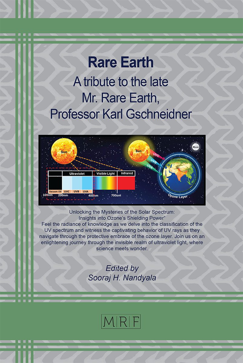
The electromagnetic spectrum of sunlight, which includes infrared (IR, 52–55%), visible (VIS, 42–43%), and ultraviolet radiation (UV, 3-5%), is a naturally occurring source of light. Out of all these radiations, UV radiations with specific wavelengths have been shown to have clinically important biological effects that are both efficient and safe for human skin. UV radiation exposure primarily aids in the formation of vitamin D synthesis and has the potential to suppress immunological response, making it a more acceptable wavelength range for the diagnosis and treatment of various skin dermatoses. While comparing natural sunlight which contains less percentage of UV radiation with broadband range artificial UV therapy is an efficient, reliable, affordable, and, nonsurgical therapeutic option available for skin dermatoses. Recent investigations and literature reviews demonstrate that various types of phototherapies such as UVB narrowband, PUVA, etc. have incompatible efficacy toward the management of dermatoses that often depend upon the action, for instance, and absorption spectra of the biological molecule (chromophores) contained in the human skin. We attempt to categorize human skin conditions by their symptoms, topological facts, and dosimetry prescribed for the cures by the minimal erythemal dosage (MED) of phototherapy recommended by the dermatologist. Descriptive analysis of types of therapies carried out on narrowband (311 ± 2 nm) and broadband (295-315 nm) ultraviolent radiation B (UVB) therapy, light-sensitive organic drug (Psoralen) in combination with UVA (PUVA) therapy and laser therapy. This review article aims at the development in the field of dermatology by reviewing historical data, therapeutic options available, a detailed understanding of skin dermatoses, and the availability of light sources for the clearance of various skin dermatoses.
Keywords
Ultraviolet Radiation, Skin Diseases, Phototherapy, Human Skin, Psoralene, Action Spectra, Absorption Spectra
Published online 6/5/2024, 34 pages
Citation: Aachal A. Sharma, D. Haranath, Overview of Treating Skin Diseases and Rejuvenating Skin using Light Sources, Materials Research Foundations, Vol. 164, pp 143-176, 2024
DOI: https://doi.org/10.21741/9781644903056-3
Part of the book on Rare Earth
References
[1] C. Karimkhani et al., “Global skin disease morbidity and mortality an update from the global burden of disease study 2013,” JAMA Dermatology, vol. 153, no. 5, pp. 406–412, 2017. https://doi.org/10.1001/jamadermatol.2016.5538
[2] A. M. Dessie, S. F. Feleke, S. G. Workie, T. G. Abebe, Y. M. Chanie, and A. K. Yalew, “Prevalence of Skin Disease and Its Associated Factors Among Primary Schoolchildren: A Cross-Sectional Study from a Northern Ethiopian Town,” Clin. Cosmet. Investig. Dermatol., vol. 15, no. April, pp. 791–801, 2022. https://doi.org/10.2147/CCID.S361051
[3] F. R. S. A. royal Harold spencer jones, “Measuring the distance of the sun from the earth,” Nature, vol. 153, pp. 181–187, 1944. https://doi.org/https://doi.org/10.1038/153181a0
[4] J. C. Zwinkels and C. Canada, “Light, Electromagnetic Spectrum,” Encycl. Color Sci. Technol., vol. 204, pp. 1–8, 2014. https://doi.org/10.1007/978-3-642-27851-8
[5] D. I. Pattison and M. J. Davies, “Actions of ultraviolet light on cellular structures.,” EXS, no. 96, pp. 131–157, 2006. https://doi.org/10.1007/3-7643-7378-4_6
[6] M. N. Mead, “Benefits of sunlight: a bright spot for human health.,” Environ. Health Perspect., vol. 116, no. 4, 2008. https://doi.org/10.1289/ehp.116-a160
[7] J. D’Orazio, S. Jarrett, A. Amaro-Ortiz, and T. Scott, “UV radiation and the skin,” Int. J. Mol. Sci., vol. 14, no. 6, pp. 12222–12248, 2013. https://doi.org/10.3390/ijms140612222
[8] C. Green, B. L. Diffey, and J. L. M. Hawk, “Ultraviolet radiation in the treatment of skin disease,” Phys. Med. Biol., vol. 37, no. 1, pp. 1–20, 1992. https://doi.org/10.1088/0031-9155/37/1/001
[9] A. L. Byrd, Y. Belkaid, and J. A. Segre, “The human skin microbiome,” Nat. Rev. Microbiol., vol. 16, no. 3, pp. 143–155, 2018. https://doi.org/10.1038/nrmicro.2017.157
[10] D. R. Bergfelt, “Anatomy and Physiology of the Mare,” Equine Breed. Manag. Artif. Insemin., pp. 113–131, 2009. https://doi.org/10.1016/B978-1-4160-5234-0.00011-8
[11] Yagi M and Yonei Y, “Glycative stress and anti-aging: 7.Glycative stress and skin aging,” Glycative Stress Res., vol. 5, no. 1, pp. 50–54, 2018
[12] S. B. Hoath and D. G. Leahy, “The Organization of Human Epidermis: Functional Epidermal Units and Phi Proportionality,” J. Invest. Dermatol., vol. 121, no. 6, pp. 1440–1446, 2003. https://doi.org/10.1046/j.1523-1747.2003.12606.x
[13] Nanda Karmaker et al., “Fundamental characteristics and application of radiation,” GSC Adv. Res. Rev., vol. 7, no. 1, pp. 064–072, 2021. https://doi.org/10.30574/gscarr.2021.7.1.0043
[14] R. P. Gallagher and T. K. Lee, “Adverse effects of ultraviolet radiation: A brief review,” Prog. Biophys. Mol. Biol., vol. 92, no. 1, pp. 119–131, 2006. https://doi.org/10.1016/j.pbiomolbio.2006.02.011
[15] “Exposure to Artificial UV Radiation and Skin Cancer,” Group, 2006
[16] K. J. Gromkowska-Kępka, A. Puścion-Jakubik, R. Markiewicz-Żukowska, and K. Socha, “The impact of ultraviolet radiation on skin photoaging — review of in vitro studies,” J. Cosmet. Dermatol., vol. 20, no. 11, pp. 3427–3431, 2021. https://doi.org/10.1111/jocd.14033
[17] R. Nair and A. Maseeh, “Vitamin D: The sunshine vitamin,” J. Pharmacol. Pharmacother., vol. 3, no. 2, pp. 118–126, 2012. https://doi.org/10.4103/0976-500X.95506
[18] M. Fors, P. González, C. Viada, K. Falcon, and S. Palacios, “Validity of the Fitzpatrick Skin Phototype Classification in Ecuador,” Adv. Ski. Wound Care, vol. 33, no. 12, pp. 1–5, 2020. https://doi.org/10.1097/01.ASW.0000721168.40561.a3
[19] V. K. Sharma, V. Gupta, B. L. Jangid, and M. Pathak, “Modification of the Fitzpatrick system of skin phototype classification for the Indian population, and its correlation with narrowband diffuse reflectance spectrophotometry,” Clin. Exp. Dermatol., vol. 43, no. 3, pp. 274–280, 2018. https://doi.org/10.1111/ced.13365
[20] V. Gupta and V. K. Sharma, “Skin typing: Fitzpatrick grading and others,” Clin. Dermatol., vol. 37, no. 5, pp. 430–436, 2019. https://doi.org/10.1016/j.clindermatol.2019.07.010
[21] A. Malerich, Sarah; Desai, “Phototherapy: A Review of Literature,” Jaocd, vol. 40, 2018
[22] H. Hönigsmann, “History of phototherapy in dermatology,” Photochem. Photobiol. Sci., vol. 12, no. 1, pp. 16–21, 2013. https://doi.org/10.1039/c2pp25120e
[23] Philips, “Effective light therapy”
[24] K. Biniek, K. Levi, and R. H. Dauskardt, “Solar UV radiation reduces the barrier function of human skin,” Proc. Natl. Acad. Sci. U. S. A., vol. 109, no. 42, pp. 17111–17116, 2012. https://doi.org/10.1073/pnas.1206851109
[25] F. Html et al., “BioMed Research International Adolescents,” vol. 2018, pp. 1–10, 2017
[26] Royal Free Hospital Department of Dermatology, “PUVA Treatment,” 2003
[27] C. W. Saleeby, “The advance of heliotherapy,” Nature, vol. 109, no. 2742, p. 663, 1922. https://doi.org/10.1038/109663a0
[28] I. G. Ferreira, M. B. Weber, and R. R. Bonamigo, “History of dermatology: the study of skin diseases over the centuries,” An. Bras. Dermatol., vol. 96, no. 3, pp. 332–345, 2021. https://doi.org/10.1016/j.abd.2020.09.006
[29] K. MCGEE, “a Synopsis of the History of Orthoptics.,” Am. Orthopt. J., vol. 14, pp. 95–98, 1964. https://doi.org/10.1080/0065955x.1965.11981449
[30] L. I. Grossweiner, The Science of Phototherapy. 2005. [Online]. Available: https://link.springer.com/book/10.1007/1-4020-2885-7#about
[31] M. Bhambri, “International Journal of Botany Studies Ammi majus: A plant with multifunctional medicinal properties,” vol. 6, no. 3, pp. 3–4, 2021, [Online]. Available: www.botanyjournals.com
[32] P. With and W. Received, “Vitiligo Photochemotherapy,” pp. 3–4, 2015
[33] A. Grzybowski, J. Sak, and J. Pawlikowski, “A brief report on the history of phototherapy,” Clin. Dermatol., vol. 34, no. 5, pp. 532–537, 2016. https://doi.org/10.1016/j.clindermatol.2016.05.002
[34] R. W. Chesney, “Theobald palm and his remarkable observation: How the sunshine vitamin came to be recognized,” Nutrients, vol. 4, no. 1, pp. 42–51, 2012. https://doi.org/10.3390/nu4010042
[35] R. Kropp, “Niels Ryberg Finsen,” Pneumologie, vol. 70, pp. S180–S183, 2016. https://doi.org/10.1055/s-0042-118376
[36] N. R. Finsen and F. Islands, “Niels Ryberg Finsen,” pp. 1–6, 1904
[37] A. hanslmeier M. vazquez, “Ultraviolet radiation in the solar system,” p. 380, 2006. https://doi.org/https://doi.org/10.1007/b13626
[38] A. E. Loignon, “Bringing Light to the World: John Harvey Kellogg and Transatlantic Light Therapy,” J. Transatl. Stud., vol. 20, no. 1, pp. 103–128, 2022. https://doi.org/10.1057/s42738-022-00092-7
[39] J. Schwarcz and D. Ph, “Colorful Nonsense : Dinshah Ghadiali and His Spectro-Chrome Device,” pp. 1–5, 2003
[40] “Parrish1974,” 2010
[41] P. S. Hemne, R. G. Kunghatkar, S. J. Dhoble, S. V. Moharil, and V. Singh, “Phosphor for phototherapy: Review on psoriasis,” Luminescence, vol. 32, no. 3, pp. 260–270, May 2017. https://doi.org/10.1002/bio.3266
[42] F. R. De Gruijl, “Biological action spectra,” Radiat. Prot. Dosimetry, vol. 91, no. 1–3, pp. 57–63, 2000. https://doi.org/10.1093/oxfordjournals.rpd.a033235
[43] G. Horneck, P. Rettberg, R. Facius, K. Scherer, and A. Medicine, “Quantification of biological effectiveness of uv radiation g. horneck, p. rettberg, r. facius and k. scherer,” vol. 9, no. Figure 1, pp. 51–52, 2006
[44] H. Hönigsmann, “Phototherapy for psoriasis,” Clin. Exp. Dermatol., vol. 26, no. 4, pp. 343–350, 2001. https://doi.org/10.1046/j.1365-2230.2001.00828.x
[45] J. P. Nater, “Phototherapy in psoriasis,” Geneesmiddelenbulletin, vol. 12, no. 9, pp. 38–41, 1978. https://doi.org/10.5005/jp/books/12839_38
[46] M. Neves-Petersen, G. Gajula, and S. Petersen, “UV Light Effects on Proteins: From Photochemistry to Nanomedicine,” Mol. Photochem. – Var. Asp., pp. 125–158, 2012
[47] N. de M. Barros, L. L. Sbroglio, M. de O. Buffara, J. L. C. e. S. Baka, A. de S. Pessoa, and L. Azulay-Abulafia, “Phototherapy,” An. Bras. Dermatol., vol. 96, no. 4, pp. 397–407, Jul. 2021. https://doi.org/10.1016/j.abd.2021.03.001
[48] R. King, K. Paver, and K. Poyzer, “Photochemotherapy in psoriasis.,” Med. J. Aust., vol. 1, no. 2, pp. 57–58, 1979. https://doi.org/10.5694/j.1326-5377.1979.tb111984.x
[49] H. Hönigsmann, “Mechanisms of phototherapy and photochemotherapy for photodermatoses,” Dermatol. Ther., vol. 16, no. 1, pp. 23–27, 2003. https://doi.org/10.1046/j.1529-8019.2003.01604.x
[50] R. B. Setlow, “Action Spectra: Skin,” Sunscreen Photobiol. Mol. Cell. Physiol. Asp., pp. 1–10, 1997. https://doi.org/10.1007/978-3-662-10135-3_1
[51] A. D. Crosswell and K. G. Lockwood, “Best practices for stress measurement: How to measure psychological stress in health research,” Heal. Psychol. Open, vol. 7, no. 2, 2020. https://doi.org/10.1177/2055102920933072
[52] B. Zhang et al., “Opportunities and Challenges: Classification of Skin Disease Based on Deep Learning,” Chinese J. Mech. Eng. (English Ed., vol. 34, no. 1, 2021. https://doi.org/10.1186/s10033-021-00629-5
[53] L. Chen et al., “Oncotarget 7204 www.impactjournals.com/oncotarget Inflammatory responses and inflammation-associated diseases in organs,” Oncotarget, vol. 9, no. 6, pp. 7204–7218, 2018, [Online]. Available: www.impactjournals.com/oncotarget/
[54] R. Parisi, I. Y. K. Iskandar, E. Kontopantelis, M. Augustin, C. E. M. Griffiths, and D. M. Ashcroft, “National, regional, and worldwide epidemiology of psoriasis: Systematic analysis and modelling study,” BMJ, vol. 369, 2020. https://doi.org/10.1136/bmj.m1590
[55] P. Hu et al., “The Role of Helper T Cells in Psoriasis,” Front. Immunol., vol. 12, no. December, pp. 1–10, 2021. https://doi.org/10.3389/fimmu.2021.788940
[56] F. O. Nestle and C. Conrad, “Mechanisms of psoriasis,” Drug Discov. Today Dis. Mech., vol. 1, no. 3, pp. 315–319, 2004. https://doi.org/10.1016/j.ddmec.2004.11.005
[57] F. Benhadou, Di. Mintoff, and V. Del Marmol, “Psoriasis: Keratinocytes or Immune Cells – Which Is the Trigger?,” Dermatology, vol. 235, no. 2, pp. 91–100, 2019. https://doi.org/10.1159/000495291
[58] S. C. Weatherhead, P. M. Farr, and N. J. Reynolds, “Spectral effects of UV on psoriasis,” Photochem. Photobiol. Sci., vol. 12, no. 1, pp. 47–53, 2013. https://doi.org/10.1039/c2pp25116g
[59] C. Ni and M. W. Chiu, “Psoriasis and comorbidities: Links and risks,” Clin. Cosmet. Investig. Dermatol., vol. 7, pp. 119–132, 2014. https://doi.org/10.2147/CCID.S44843
[60] C. Balakrishnan and N. Madnani, “Diagnosis and management of psoriatic arthritis,” Indian J. Dermatol. Venereol. Leprol., vol. 79, no. SUPPL. 1, pp. 18–24, 2013. https://doi.org/10.4103/0378-6323.115507
[61] C. Niu, X. Lu, and H. A. Aisa, “Preparation of novel 1,2,3-triazole furocoumarin derivatives via click chemistry and their anti-vitiligo activity,” RSC Adv., vol. 9, no. 3, pp. 1671–1678, 2019. https://doi.org/10.1039/C8RA09755K
[62] A. Arora and M. Kumaran, “Pathogenesis of vitiligo: An update,” Pigment Int., vol. 4, no. 2, p. 65, 2017. https://doi.org/10.4103/2349-5847.219673
[63] M. L. Frisoli, K. Essien, and J. E. Harris, “Vitiligo: Mechanisms of Pathogenesis and Treatment,” Annu. Rev. Immunol., vol. 38, pp. 621–648, 2020. https://doi.org/10.1146/annurev-immunol-100919-023531
[64] C. E. Artz, C. M. Farmer, and H. W. Lim, “Polymorphous Light Eruption: a Review,” Curr. Dermatol. Rep., vol. 8, no. 3, pp. 110–116, 2019. https://doi.org/10.1007/s13671-019-0264-y
[65] I. An, M. Harman, and I. Ibiloglu, “Polymorphus Light Eruption-An Indian Scenario,” Indian Dermatol. Online J., vol. 10, no. 4, pp. 481–485, 2017. https://doi.org/10.4103/idoj.IDOJ
[66] S. E. Ullrich, “Does exposure to UV radiation induce a shift to a Th-2-like immune reaction?,” Photochem. Photobiol., vol. 64, no. 2, pp. 254–258, 1996. https://doi.org/10.1111/j.1751-1097.1996.tb02454.x
[67] E. Vicenzi and G. Poli, “Ultraviolet irradiation and cytokines as regulators of HIV latency and expression,” Chem. Biol. Interact., vol. 91, no. 2–3, pp. 101–109, 1994. https://doi.org/10.1016/0009-2797(94)90030-2
[68] H. W. Lim, S. Vallurupalli, T. Meola, and N. A. Soter, “UVB phototherapy is an effective treatment for pruritus in patients infected with HIV,” J. Am. Acad. Dermatol., vol. 37, no. 3 I, pp. 414–417, 1997. https://doi.org/10.1016/S0190-9622(97)70142-7
[69] A. Alothman and H. S. H. Mohamed, “Recurrent Nasal Sebaceous Carcinoma in Human Immunodefiency Virus / Hepatitis B Virus ( HIV / HBV ) Coinfection Patient : A Case Report,” vol. 3, no. 10, pp. 329–332, 2015. https://doi.org/10.12691/ajmcr-3-10-7
[70] T. Meola, N. A. Soter, R. Ostreicher, M. Sanchez, and J. A. Moy, “The safety of UVB phototherapy in patients with HIV infection,” J. Am. Acad. Dermatol., vol. 29, no. 2, pp. 216–220, 1993. https://doi.org/10.1016/0190-9622(93)70171-O
[71] S. Mattinen, A. Lagerstedt, and K. Krohn, “Effect of PUVA on immunologic and virologic findings in HIV-infected patients,” J. Am. Acad. Dermatol., vol. 24, no. 3, pp. 404–410, 1991. https://doi.org/10.1016/0190-9622(91)70060-F
[72] J. M. Nuhu, J. Mohammed, and M. Muhammad, “UV therapy: Physiotherapists’ perception of therapeutic efficacy and barriers to usage,” Hong Kong Physiother. J., vol. 32, no. 1, pp. 44–48, 2014. https://doi.org/10.1016/j.hkpj.2014.02.002
[73] R. P. Rastogi, Richa, A. Kumar, M. B. Tyagi, and R. P. Sinha, “Molecular mechanisms of ultraviolet radiation-induced DNA damage and repair,” J. Nucleic Acids, vol. 2010, 2010. https://doi.org/10.4061/2010/592980
[74] T. M. Lotti and S. Gianfaldoni, “Ultraviolet A-1 in dermatological diseases,” Adv. Exp. Med. Biol., vol. 996, pp. 105–110, 2017. https://doi.org/10.1007/978-3-319-56017-5_9
[75] F. Bernerd, T. Passeron, I. Castiel, and C. Marionnet, “The Damaging Effects of Long UVA (UVA1) Rays: A Major Challenge to Preserve Skin Health and Integrity,” Int. J. Mol. Sci., vol. 23, no. 15, pp. 1–33, 2022. https://doi.org/10.3390/ijms23158243
[76] D. L. Damian, Y. J. Matthews, T. A. Phan, and G. M. Halliday, “An action spectrum for ultraviolet radiation-induced immunosuppression in humans,” Br. J. Dermatol., vol. 164, no. 3, pp. 657–659, 2011. https://doi.org/10.1111/j.1365-2133.2010.10161.x
[77] D. D. Moyal and A. M. Fourtanier, “Effects of UVA radiation on an established immune response in humans and sunscreen efficacy,” Exp. Dermatology, Suppl., vol. 11, no. 1, pp. 28–32, 2002. https://doi.org/10.1034/j.1600-0625.11.s.1.7.x
[78] I. Furocoumarins, “Yearly Review Recent Advances in Psoralen Phototoxicity,” vol. 50, no. 6, pp. 859–882, 1989
[79] R. Vangipuram and S. R. Feldman, “Ultraviolet phototherapy for cutaneous diseases: A concise review,” Oral Dis., vol. 22, no. 4, pp. 253–259, 2016. https://doi.org/10.1111/odi.12366
[80] J. L. Bolognia, “Rectal suppositories of 8-methoxsalen produce fewer gastrointestinal side effects than the oral formulation,” J. Am. Acad. Dermatol., vol. 35, no. 3 PART I, pp. 424–427, 1996. https://doi.org/10.1016/s0190-9622(96)90609-x
[81] J. Foerster et al., “Narrowband UVB treatment is highly effective and causes a strong reduction in the use of steroid and other creams in psoriasis patients in clinical practice,” PLoS One, vol. 12, no. 8, pp. 1–14, 2017. https://doi.org/10.1371/journal.pone.0181813
[82] K. Boswell et al., “Narrowband ultraviolet B treatment for psoriasis is highly economical and causes significant savings in cost for topical treatments,” Br. J. Dermatol., vol. 179, no. 5, pp. 1148–1156, 2018. https://doi.org/10.1111/bjd.16716
[83] P. V Prasad, T. Paari, K. Chokkalingam, and V. Vijaybushanam, “Malignant syphilis (leus maligna) in a HIV infected patient.,” Indian journal of dermatology, venereology and leprology, vol. 67, no. 4. pp. 192–4, 2001. [Online]. Available: https://www.ncbi.nlm.nih.gov/pubmed/17664738
[84] D. Bilsland et al., “An appraisal of narrowband (TL-01) UVB phototherapy. British Photodermatology Group Workshop Report (April 1996),” Br. J. Dermatol., vol. 137, no. 3, pp. 327–330, 1997. https://doi.org/10.1111/j.1365-2133.1997.tb03733.x
[85] S. H. Ibbotson et al., “An update and guidance on narrowband ultraviolet B phototherapy: A British Photodermatology Group Workshop Report,” Br. J. Dermatol., vol. 151, no. 2, pp. 283–297, 2004. https://doi.org/10.1111/j.1365-2133.2004.06128.x
[86] K. Ly, M. P. Smith, Q. G. Thibodeaux, K. M. Beck, W. Liao, and T. Bhutani, “Beyond the Booth: Excimer Laser for Cutaneous Conditions,” Dermatol. Clin., vol. 38, no. 1, pp. 157–163, 2020. https://doi.org/10.1016/j.det.2019.08.009
[87] T. B. M. Abrounk, E. Levin, M. Brodsky, J. R. Gandy, “Excimer laser for the treatment of psoriasis: safety, efficacy, and patient acceptibility,” Psoriasistarget Ther., no. 6, pp. 165–173, 2016
[88] C. Lopes, V. F. M. Trevisani, and T. Melnik, “Efficacy and Safety of 308-nm Monochromatic Excimer Lamp Versus Other Phototherapy Devices for Vitiligo: A Systematic Review with Meta-Analysis,” Am. J. Clin. Dermatol., vol. 17, no. 1, pp. 23–32, 2016. https://doi.org/10.1007/s40257-015-0164-2
[89] C. A. Robertson, D. H. Evans, and H. Abrahamse, “Photodynamic therapy (PDT): A short review on cellular mechanisms and cancer research applications for PDT,” J. Photochem. Photobiol. B Biol., vol. 96, no. 1, pp. 1–8, 2009. https://doi.org/10.1016/j.jphotobiol.2009.04.001
[90] S. Kwiatkowski et al., “Photodynamic therapy – mechanisms, photosensitizers and combinations,” Biomed. Pharmacother., vol. 106, no. July, pp. 1098–1107, 2018. https://doi.org/10.1016/j.biopha.2018.07.049
[91] M. Nakamura, B. Farahnik, and T. Bhutani, “Recent advances in phototherapy for psoriasis [version 1; referees: 2 approved],” F1000Research, vol. 5, pp. 1–8, 2016. https://doi.org/10.12688/F1000RESEARCH.8846.1
[92] A. Carolina Sparavigna, “Carbon-Arc Light as the Electric Light of 1870,” Int. J. Sci., vol. 0, no. 10, pp. 1–7, 2014. https://doi.org/10.18483/ijsci.581
[93] R. Jenkins, B. Aldwell, S. Yin, M. Meyer, A. J. Robinson, and R. Lupoi, “Energy efficiency of a quartz tungsten halogen lamp: Experimental and numerical approach,” Therm. Sci. Eng. Prog., vol. 13, no. July, p. 100385, 2019. https://doi.org/10.1016/j.tsep.2019.100385
[94] A. Namin, C. Jivacate, D. Chenvidhya, K. Kirtikara, and J. Thongpron, “Construction of tungsten halogen, pulsed LED, and combined tungsten halogen-LED solar simulators for solar cell i – V characterization and electrical parameters determination,” Int. J. Photoenergy, vol. 2012, 2012. https://doi.org/10.1155/2012/527820
[95] H. Kohli, S. Srivastava, S. K. Sharma, S. Chouhan, and M. Oza, “Design of Programmable LED Based Phototherapy System,” Int. J. Opt., vol. 2019, 2019. https://doi.org/10.1155/2019/6023646
[96] H. J. Vreman, R. J. Wong, and D. K. Stevenson, “Phototherapy: Current methods and future directions,” Semin. Perinatol., vol. 28, no. 5, pp. 326–333, Oct. 2004. https://doi.org/10.1053/j.semperi.2004.09.003
[97] Beggs, Sarah BS; Short, Jack MD; Rengifo-Pardo, Monica MD; Ehrlich, Alison MD, MHS, “Applications of the Excimer Laser: A Review”,”American Society for Dermatologic Surgery”.,vol. 41, pp. 1201–1211, 2015. https://doi.org/10.1097/DSS.0000000000000485